. 2012 Dec;139(24):4666-74.
doi: 10.1242/dev.066647.

Macrophage migration inhibitory factor acts as a neurotrophin in the developing inner ear

Affiliations

Macrophage migration inhibitory factor acts as a neurotrophin in the developing inner ear

Lisa M Bank et al. Development. 2012 Dec.

Erratum in

  • Development. 2013 Jan 15;140(2):479. Koch, Alicia E [corrected to Koch, Alisa E]

Abstract

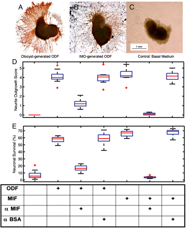
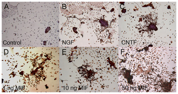
This study is the first to demonstrate that macrophage migration inhibitory factor (MIF), an immune system 'inflammatory' cytokine that is released by the developing otocyst, plays a role in regulating early innervation of the mouse and chick inner ear. We demonstrate that MIF is a major bioactive component of the previously uncharacterized otocyst-derived factor, which directs initial neurite outgrowth from the statoacoustic ganglion (SAG) to the developing inner ear. Recombinant MIF acts as a neurotrophin in promoting both SAG directional neurite outgrowth and neuronal survival and is expressed in both the developing and mature inner ear of chick and mouse. A MIF receptor, CD74, is found on both embryonic SAG neurons and adult mouse spiral ganglion neurons. Mif knockout mice are hearing impaired and demonstrate altered innervation to the organ of Corti, as well as fewer sensory hair cells. Furthermore, mouse embryonic stem cells become neuron-like when exposed to picomolar levels of MIF, suggesting the general importance of this cytokine in neural development.


Figures

Fig. 1.
Fig. 1.
Effect of treating MIF or ODF used in assays of SAG neurite outgrowth and neuron survival with MIF function-blocking antibody. (A-C) SAG neurite outgrowth is abundant after 24 hours of exposure to Immortomouse otocyst (IMO)-generated ODF (A) or to E5 chick otocyst-generated ODF (B). No neurite outgrowth is seen in basal serum-free medium (C). (D) Similar neurite outgrowth was promoted by IMO-generated ODF and 10 ng/ml recombinant MIF (rMIF), but not by control medium (measured on a scale of 1-5; the chart at the bottom indicates treatment conditions for both D and E). Pretreatment of rMIF with a MIF function-blocking antibody (α MIF) substantially inhibited the neurite outgrowth-promoting activity of MIF, whereas pretreatment with anti-BSA antibody (α BSA) did not. (E) Neuronal survival. The anti-MIF antibody treatment reduced neuronal survival to background levels when added to rMIF, but not to ODF, indicating that there is residual neuron survival-promoting activity that is not attributable to MIF. Neurite outgrowth: ODF alone, n=15 replicates; ODF + anti-MIF, n=7; ODF + anti-BSA, n=8; MIF alone, n=8; MIF + anti-MIF, n=10; MIF + anti-BSA, n=8. Neuronal survival: ODF alone, n=12; ODF + anti-MIF, n=8; ODF + anti-BSA, n=7; MIF alone, n=9; MIF + anti-MIF, n=8; MIF + anti-BSA, n=8. See supplementary material Table S1 for statistical significance.
Fig. 2.
Fig. 2.
mESCs treated with MIF take on a neuron-like morphology. (A) D3 mESCs grown in control cultures with no growth factors added did not become neuron-like. (B,C) By contrast, D3 mESCs treated with the growth factors NGF (B) or CNTF (C) produced neuron-like cells, with neurofilament-positive processes. (D-F) Recombinant mouse MIF (rmMIF) at 1 ng/ml (D), 10 ng/ml (E) and 50 ng/ml (F) also produced neuron-like cells with elaborate neurofilament-positive processes seen as early as 48 hours in culture. Both lower (1 pg/ml) and higher (500 ng/ml) concentrations of rmMIF were also effective (not shown). Phase contrast image (magnification 4×).
Fig. 3.
Fig. 3.
MIF protein is widely expressed in E9.5 mouse otocysts and hindbrain. MIF labeling in hindbrain (NT) is ventral and that in the otocysts (O) is medial on the hindbrain side. Epithelial cells lining the otocyst, particularly at the dorsal edge, are labeled, as are epithelial cells on the external medial side of the otocyst and cells in the otocyst medial wall. Inset shows a control without primary antibody.
Fig. 4.
Fig. 4.
Expression of MIF and MCP1 proteins in the cochlea of WT Balb/c mice. Cryosections of Balb/c cochleae were labeled for MIF (green) and MCP1 (red). MIF is expressed in SL, SpL, StV, SP and SCs. MCP1 expression is found in IHCs and OHCs, as well as in StV and basement membranes. The region of colocalization (yellow) indicates the portion of the SCs that directly contact and cup and the IHC and OHC. IHC, inner hair cell; OHC, outer hair cell; RM, Reissner’s membrane; SM, scala media; SV, scala vestibuli; SL, spiral ligament; SpL, spiral limbus; SP, spiral prominences; SC, supporting cell; StV, stria vascularis; TM, tectorial membrane. Magnification 20×.
Fig. 5.
Fig. 5.
Characterization of Mif KO embryonic and adult inner ear structures. (A-F) Epifluorescence of organs of Corti (OC) labeled for neurofilament (a neuronal marker, red) and myosin VIIa (an HC marker, green) in WT (Balb/c) (A-C) and Mif KO (D-F) mice. (A-C) Both IHCs and OHCs in the cochlea are phenotypically normal in the OC in WT mice. (D-F) There is a lower neuron density (D,F) and areas of missing HCs (E,F) in the Mif KO OC. Myosin labeling is considerably reduced in the three rows of outer HCs as well as in the one row of inner HCs in the KO animal (E,F). (G,H) Low-magnification view of the basal 48 kHz region of the cochlea of WT (G) and Mif KO (H) animals. Note that, as in B and C, there is evidence of green fluorescence (myosin VIIa labeling) in the WT OC and also some evidence of labeling in the row of IHCs in the Mif KO animal; myosin VIIa labeling is not apparent in the three rows of WT OHCs (G), where the DAPI labeling (nuclear marker, blue) becomes apparent. Neurofilament is in red. Both missing HCs and loss of myosin VIIa label are apparent in the Mif KO animal (H). Arrow (H) indicates an area with large numbers of missing HCs.
Fig. 6.
Fig. 6.
Neurites from WT SG extend towards WT but not towards Mif KO OC. Spiral ganglia (SG) from WT (Balb/c) mice were excised at P3 and placed in organ culture with OC from either WT (A) or Mif KO (B) P3 mice. (A) After 5 days in culture, there was robust and directional neurite outgrowth from the SG towards the WT OC. Note that the trajectory of the neuronal processes is always towards the WT OC (yellow arrows). In every co-culture (n=19), neurites from the SG made contact with the explanted WT OC. (B) By contrast, when the WT SG was placed in culture with the Mif KO OC, neurites extended radially and uniformly for only short distances. None of the neurites made contact with the Mif KO OC in any experiment (n=17). o of C, organ of Corti; SGN, spiral ganglion neurons from SG explant.

Similar articles

Cited by

References

    1. Armstrong B. D., Hu Z., Abad C., Yamamoto M., Rodriguez W. I., Cheng J., Tam J., Gomariz R. P., Patterson P. H., Waschek J. A. (2003). Lymphocyte regulation of neuropeptide gene expression after neuronal injury. J. Neurosci. Res. 74, 240–247 - PubMed
    1. Bajetto A., Bonavia R., Barbero S., Florio T., Schettini G. (2001). Chemokines and their receptors in the central nervous system. Front. Neuroendocrinol. 22, 147–184 - PubMed
    1. Barald K. F., Kelley M. W. (2004). From placode to polarization: new tunes in inner ear development. Development 131, 4119–4130 - PubMed
    1. Barald K. F., Lindberg K. H., Hardiman K., Kavka A. I., Lewis J. E., Victor J. C., Gardner C. A., Poniatowski A. (1997). Immortalized cell lines from embryonic avian and murine otocysts: tools for molecular studies of the developing inner ear. Int. J. Dev. Neurosci. 15, 523–540 - PubMed
    1. Bernhagen J., Krohn R., Lue H., Gregory J. L., Zernecke A., Koenen R. R., Dewor M., Georgiev I., Schober A., Leng L., et al. (2007). MIF is a noncognate ligand of CXC chemokine receptors in inflammatory and atherogenic cell recruitment. Nat. Med. 13, 587–596 - PubMed

MeSH terms

Substances