. 2012;7(11):e48871.
doi: 10.1371/journal.pone.0048871. Epub 2012 Nov 16.

Osteoclasts in multiple myeloma are derived from Gr-1+CD11b+myeloid-derived suppressor cells

Affiliations

Osteoclasts in multiple myeloma are derived from Gr-1+CD11b+myeloid-derived suppressor cells

Junling Zhuang et al. PLoS One. 2012.

Abstract

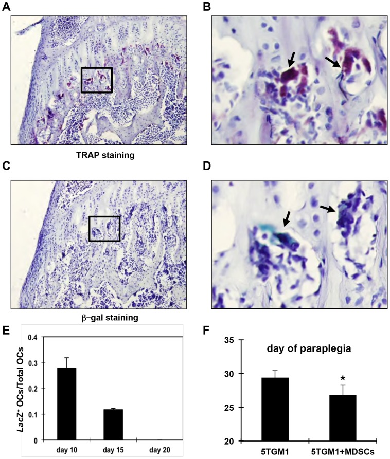
Osteoclasts play a key role in the development of cancer-associated osteolytic lesions. The number and activity of osteoclasts are often enhanced by tumors. However, the origin of osteoclasts is unknown. Myeloid-derived suppressor cells (MDSCs) are one of the pre-metastatic niche components that are induced to expand by tumor cells. Here we show that the MDSCs can differentiate into mature and functional osteoclasts in vitro and in vivo. Inoculation of 5TGM1-GFP myeloma cells into C57BL6/KaLwRij mice led to a significant expansion of MDSCs in blood, spleen, and bone marrow over time. When grown in osteoclastogenic media in vitro, MDSCs from tumor-challenged mice displayed 14 times greater potential to differentiate into mature and functional osteoclasts than those from non-tumor controls. Importantly, MDSCs from tumor-challenged LacZ transgenic mice differentiated into LacZ+osteoclasts in vivo. Furthermore, a significant increase in tumor burden and bone loss accompanied by increased number of osteoclasts was observed in mice co-inoculated with tumor-challenged MDSCs and 5TGM1 cells compared to the control animals received 5TGM1 cells alone. Finally, treatment of MDSCs from myeloma-challenged mice with Zoledronic acid (ZA), a potent inhibitor of bone resorption, inhibited the number of osteoclasts formed in MDSC cultures and the expansion of MDSCs and bone lesions in mice. Collectively, these data provide in vitro and in vivo evidence that tumor-induced MDSCs exacerbate cancer-associated bone destruction by directly serving as osteoclast precursors.


Conflict of interest statement

Competing Interests: The authors have declared that no competing interests exist.

Figures

Figure 1
Figure 1. Myeloma increased Gr-1+CD11b+cells in mice. A–B,
Flow cytometry analysis showing the percentage of the Gr-1+CD11b+cells in bone marrow (A) and spleen (B) of control and 5TGM1-GFP inoculated mice 28 days after tumor inoculation. C–D, The quantification of flow cytometry of A–B. *P<0.05, n = 6. C∼D, Myeloma model validation. Percentage of 5TGM1-GFP+myeloma cells (C) and serum IgG2bκ level (mg/ml) (B) in C57BL6/KaLwRij mice increased with time after 5TGM1 myeloma cell inoculation. n = 6 for tumor group; n = 4 for control group.
Figure 2
Figure 2. Tumor-associated MDSCs are osteoclast precursors.
A. Gr-1 expression decreases with cultured time in MDSCs from myeloma-bearing mice. Note that FACS sorted MDSCs were double positive for both Gr-1 and CD11b surface markers (98%) at the beginning of the culture time. However, the percentage of Gr-1-expressing cells progressively dropped with cultured time. B–D. Immunocytochemistry using indicated antibodies. IgG, negative control (B); αCRLR, Calcitonin receptor-like receptor (C); αCD51 (VNRαv chain. EF, TRAP staining of MDSC cultures of nontumor-bearing mice (control, E) or tumor-bearing mice (5TGM1, F) grown under osteoclastogenic condition for 14 days. G. Quantification of E and F. H. Dentine assays demonstrating that osteoclasts derived from MDSCs of tumor-bearing mice has greater activity to resorb bone.
Figure 3
Figure 3. Gr-1+CD11b+cells from Rag2 −/− myeloma-bearing mice form osteoclasts. A,
Flow cytometry graph showing isolated Gr-1+CD11b+cells from 5TGM1-challenged mice. B, Quantification of multinucleated TRAP+cell number (# OC) of Gr-1+CD11b+cultures from nontumor-bearing control (control) or 5TMG1-challenged mice (5TGM1). Cells were cultured for 14 days under osteoclast differentiation condition. C–D, TRAP staining of Gr-1+CD11b+cultures from control or myeloma-bearing Rag2 −/− mice (100×).
Figure 4
Figure 4. Gr-1+CD11b+cells from myeloma-bearing mice differentiate into osteoclasts in vivo. A–B.
β-galactosidase staining of bone sections of tumor-bearing mice 10 days after co-inoculation of 5TGM1 cells and Gr-1+CD11b+cells of tumor-challenged LacZ;Rag2 −/− mice. Note the multinucleated LacZ+cells (blue) present under the growth plate in A (200×) and B (400× of the square in A). B–C. TRAP staining showing multinucleated osteoclasts. C, 200×; D, 400× of indicated area in C. E, The ratio of LacZ+osteoclasts over the total number of multinucleated TRAP+osteoclasts at indicated days after coinjection of 5TGM1 and MDSCs of myeloma-challenged LacZTg;Rag2−/− mice. n = 3. F. Tumor-associated MDSCs facilitate paraplegia in 5TGM1-induced paraplegia in mice. *p = 0.02, n = 6 for tumor group; n = 5 for control group.
Figure 5
Figure 5. Gr-1+CD11b+cells from myeloma-bearing mice increased tumor burden and bone lesion. A.
Quantification of GFP+5TGM1 cells in bone marrow and spleen of mice after co-inoculation of 5TGM1-GFP and MDSCs of myeloma-challenged or inoculation of 5TGM1 alone. B. Bone lesion assessed by quantification of bone volume over total volume (BV/TV) on histology of tibial sections. N.S., not significant. C,TRAP staining of tibial sections (100x) and D, quantification of OC #/bone surface showing the increased osteoclast number in 5TGM1 only and 5TGM1+Tu MDSCs groups compared with non-tumor control. Note that osteoclast numbers increased in mice coinoculated with MDSCs and 5TGM1 compared to mice inoculated with 5TGM1 alone.
Figure 6
Figure 6. Zoledronic acid (ZA) inhibits osteoclast differentiation of myeloma-induced+MDSCs in vitro. A–E,
Photographic images of the cell cultures of Gr-1+CD11b+cells isolated from spleen of myeloma-bearing mice. Cells were treated with ZA at indicated concentrations and TRAP staining was performed at day 14 post osteoclastogenic induction. F. Multinucleated cells were quantified as the number of multunucleated TRAP+osteoclasts per 100× field. n = 3.
Figure 7
Figure 7. Zoledronic acid (ZA) decreases the myeloma-induced expansion of Gr-1+CD11b+cells in vivo. A.
FACS analysis showing the percentage of Gr-1+CD11b+cells in spleen of control, tumor-bearing mice that were non-treated (5TGM1) and treated with ZA (5TGM1+ZA). B, TRAP staining of tibial bone sections from tumor-bearing mice that were non-treated (5TGM1) or treated with ZA (5TGM1+ZA). C, Quantification of osteoclast number over bone surface of B. *p<0.05, n = 5 for each group.
Figure 8
Figure 8. ZA inhibits osteoclast differentiation of Gr-1+CD11b+cells via a prenylation-independent manner. A.
TRAP staining of Gr-1+CD11b+cultures of tumor-bearing mice nontreated (5TGM1) or treated with ZA under osteoclast differentiation medium for 14 days (100×). B. Quantification of osteoclast number of A. C. Western blot showing that in vivo treatment of ZA does not affect prenylation of Rap1A and Rab6 in MDSCs of tumor-bearing mice. Note that prenylation of Rap1A and Rab6 were inhibited by ZA treatment in 5TGM1 cells in vitro.

References

    1. Kaplan RN, Rafii S, Lyden D (2006) Preparing the “soil”: the premetastatic niche. Cancer research 66: 11089–11093. - PMC - PubMed
    1. Kaplan RN, Psaila B, Lyden D (2007) Niche-to-niche migration of bone-marrow-derived cells. Trends in molecular medicine 13: 72–81. - PubMed
    1. Kaplan RN, Psaila B, Lyden D (2006) Bone marrow cells in the ‘pre-metastatic niche’: within bone and beyond. Cancer metastasis reviews 25: 521–529. - PubMed
    1. Psaila B, Kaplan RN, Port ER, Lyden D (2006) Priming the ‘soil’ for breast cancer metastasis: the pre-metastatic niche. Breast disease 26: 65–74. - PubMed
    1. Edwards CM, Zhuang J, Mundy GR (2008) The pathogenesis of the bone disease of multiple myeloma. Bone 42: 1007–1013. - PMC - PubMed

Publication types

MeSH terms