. 2012 Nov 21;3(11):963-71.
doi: 10.1021/cn300110c. Epub 2012 Sep 24.

Glycogen synthase kinase 3 inhibition promotes adult hippocampal neurogenesis in vitro and in vivo

Affiliations

Glycogen synthase kinase 3 inhibition promotes adult hippocampal neurogenesis in vitro and in vivo

Jose A Morales-Garcia et al. ACS Chem Neurosci. .

Abstract

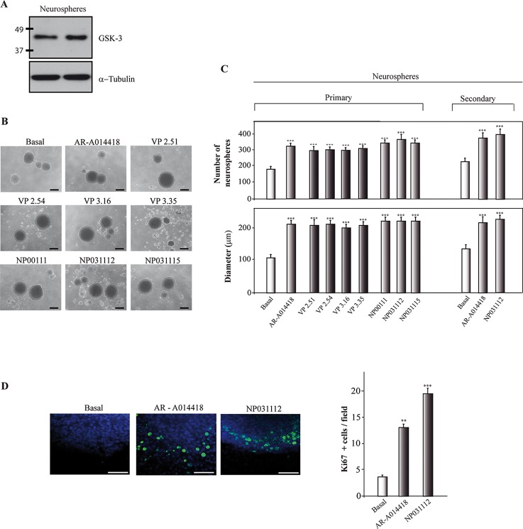
Glycogen synthase kinase-3 (GSK-3) is a serine/threonine kinase originally identified as a regulator of glycogen metabolism but it also plays a pivotal role in numerous cellular functions, including differentiation, cell cycle regulation, and proliferation. The dentate gyrus of the hippocampus, together with the subventricular zone of the lateral ventricles, is one of the regions in which neurogenesis takes place in the adult brain. Here, using a chemical genetic approach that involves the use of several diverse inhibitors of GSK-3 as pharmacological tools, we show that inhibition of GSK-3 induces proliferation, migration, and differentiation of neural stem cells toward a neuronal phenotype in in vitro studies. Also, we demonstrate that inhibition of GSK-3 with the small molecule NP03112, called tideglusib, induces neurogenesis in the dentate gyrus of the hippocampus of adult rats. Taken together, our results suggest that GSK-3 should be considered as a new target molecule for modulating the production and integration of new neurons in the hippocampus as a treatment for neurodegenerative diseases or brain injury and, consequently, its inhibitors may represent new potential therapeutic drugs in neuroregenerative medicine.


Figures

Figure 1
Figure 1
Chemical structures, IC50 values regarding GSK-3β inhibition, and binding mode of the small heterocycles GSK-3 inhibitors used as molecular probes.
Figure 2
Figure 2
Effects of GSK-3 inhibitors on neurosphere formation. (A) Representative Western blot showing expression of GSK-3 in neurospheres derived from hippocampus. (B)Representative phase-contrast micrographs showing the size of primary neurospheres after 7 days in culture in the presence or absence of the different inhibitors. Scale bar = 50 μm. (C) Quantification analysis of the primary and secondary neurosphere number. The number of neurospheres was counted and their diameter measured. The diameter of at least 50 neurospheres and the total number of neurospheres was determined in control and GSK-3 inhibitor-treated cultures. Results are mean values ± SD from three independent experiments. **P ≤ 0.01; ***p ≤ 0.001. (D) Representative confocal images of Ki67 immunoreactivity (green) in primary neurospheres. DAPI staining (blue) was used as a nuclear marker. Quantification of Ki67-positive cells is shown. Results are mean values ± SD from three independent experiments performed in triplicate. **P ≤ 0.01; ***P ≤ 0.001. Scale bar = 50 μm.
Figure 3
Figure 3
Effects of GSK-3β inhibitors on cell migration out of the neurospheres. (A) Single neurospheres were plated on polylysine-coated coated culture dishes in the presence or absence of the inhibitors and the cell migration out of the sphere was monitored 24 h later. Representative photomicrographs are shown. Scale bars = 50 μm. (B) Quantitative data of the furthest distance of cell migration. **P ≤ 0.01; ***P ≤ 0.001.
Figure 4
Figure 4
Effects of GSK-3 inhibitors on neural stem cell differentiation. The neurospheres were grown for 7 days in the presence or absence of GSK-3 inhibitors and then adhered for 2 days to allow differentiation. Neuronal cells were detected using an anti-β-tubulin antibody (TuJ clone, red) and astrocytes using an anti-GFAP (green) antibody. DAPI marker (blue) was used for nuclear staining. Scale bar = 30 μm.
Figure 5
Figure 5
Effects of GSK-3 inhibitors on proliferating cells in the hippocampus. (A) Representative coronal sections showing BrdU-labeled cells in the hippocampus. Scale bar = 100 μm. Insets show higher magnifications of representative areas in the dentate gyrus. Scale bar = 50 um. (B) Quantification of BrdU positive cells in the hippocampus. Values are the mean ± SD from five different animals. ***P ≤ 0.05.
Figure 6
Figure 6
Effects of GSK-3 inhibitors on neuronal differentiation of hippocampal neural stem cells. Representative coronal sections of the hippocampus showing the expression of DCX (A) and PSA-NCAM (B) in the subgranular zone of the dentate gyrus (DG). Scale bar = 100 μm. Insets show higher magnification of the DG. Scale bar = 50 μm.

Similar articles

Cited by

References

    1. Gage F. H. (2000) Mammalian neural stem cells. Science 287, 1433–1438. - PubMed
    1. Gage F. H.; Kempermann G.; Palmer T. D.; Peterson D. A.; Ray J. (1998) Multipotent progenitor cells in the adult dentate gyrus. J. Neurobiol. 36, 249–266. - PubMed
    1. Eriksson P. S.; Perfilieva E.; Bjork-Eriksson T.; Alborn A. M.; Nordborg C.; Peterson D. A.; Gage F. H. (1998) Neurogenesis in the adult human hippocampus. Nat. Med. 4, 1313–1317. - PubMed
    1. Kee N.; Teixeira C. M.; Wang A. H.; Frankland P. W. (2007) Preferential incorporation of adult-generated granule cells into spatial memory networks in the dentate gyrus. Nat. Neurosci. 10, 355–362. - PubMed
    1. Zhao C.; Teng E. M.; Summers R. G. Jr.; Ming G. L.; Gage F. H. (2006) Distinct morphological stages of dentate granule neuron maturation in the adult mouse hippocampus. J. Neurosci. 26, 3–11. - PMC - PubMed

Publication types