. 2012 Nov 23:13:662.
doi: 10.1186/1471-2164-13-662.

Identification of novel genes involved in DNA damage response by screening a genome-wide Schizosaccharomyces pombe deletion library

Affiliations

Identification of novel genes involved in DNA damage response by screening a genome-wide Schizosaccharomyces pombe deletion library

Xian Pan et al. BMC Genomics. .

Abstract

Background: DNA damage response (DDR) plays pivotal roles in maintaining genome integrity and stability. An effective DDR requires the involvement of hundreds of genes that compose a complicated network. Because DDR is highly conserved in evolution, studies in lower eukaryotes can provide valuable information to elucidate the mechanism in higher organisms. Fission yeast (Schizosaccharomyces pombe) has emerged as an excellent model for DDR research in recent years. To identify novel genes involved in DDR, we screened a genome-wide S. pombe haploid deletion library against six different DNA damage reagents. The library covered 90.5% of the nonessential genes of S. pombe.

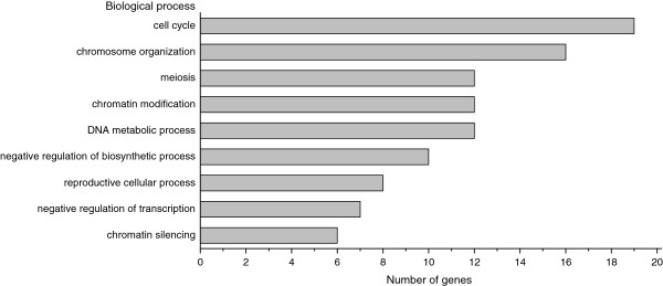
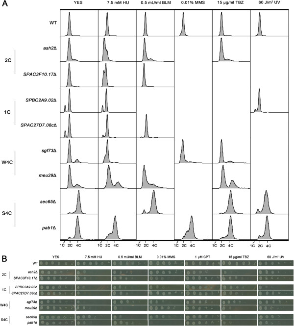
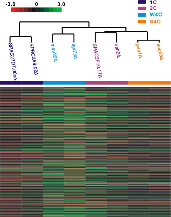
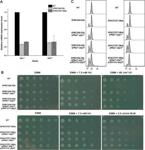
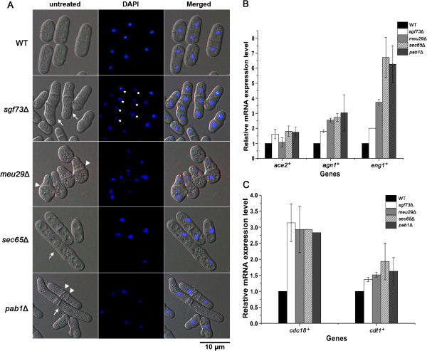
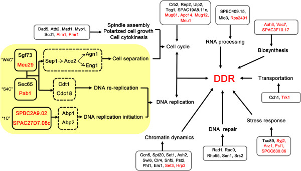
Results: We have identified 52 genes that were actively involved in DDR. Among the 52 genes, 20 genes were linked to DDR for the first time. Flow cytometry analysis of the repair defective mutants revealed that most of them exhibited a defect in cell cycle progression, and some caused genome instability. Microarray analysis and genetic complementation assays were carried out to characterize 6 of the novel DDR genes in more detail. Data suggested that SPBC2A9.02 and SPAC27D7.08c were required for efficient DNA replication initiation because they interacted genetically with DNA replication initiation proteins Abp1 and Abp2. In addition, deletion of sgf73+, meu29+, sec65+ or pab1+ caused improper cytokinesis and DNA re-replication, which contributed to the diploidization in the mutants.

Conclusions: A genome-wide screen of genes involved in DDR emphasized the key role of cell cycle control in the DDR network. Characterization of novel genes identified in the screen helps to elucidate the mechanism of the DDR network and provides valuable clues for understanding genome stability in higher eukaryotes.


Figures

Figure 1
Figure 1
Representative GO terms of 52 genes. GO terms shown in the chart are: cell cycle (GO:0007049), chromosome organization (GO:0051276), meiosis (GO:0007126), chromatin modification (GO:0016568), DNA metabolic process (GO:0006259), negative regulation of biosynthetic process (GO:0009890), reproductive cellular process (GO:0048610), negative regulation of transcription (GO:0016481) and chromatin silencing (GO:0006342). Complete list of GO terms is shown in Additional file 1: Table S2.
Figure 2
Figure 2
Flow cytometry analysis and spot assays of eight representative mutants. (A) Flow cytometry analysis of eight mutants. Cells were grown to the logarithmic phase and treated with a DNA damage reagent for 2 h. For UV sensitivity assay, cells were exposed to 60 J/m2 radiation and then grown for 2 h. After treatment cells were harvested and subjected to cytometry analysis. (B) Sensitivity to different DNA damage reagents was quantified by spot assays. Exponentially growing cells, WT or deletions, were harvested and 5-fold serial dilutions were spotted on plates supplemented with DNA damage reagents. The plates were photographed after 3~4 days of incubation at 32°C.
Figure 3
Figure 3
Clustering analysis of eight mutants. Hierarchical clustering matrix of eight mutants with microarray data for S pombe gene transcription. Color panel indicates relative increase (red) and decrease (green) to the median (black) of eight mutants for each transcript.
Figure 4
Figure 4
abp1+ and abp2+ function downstream of SPBC2A9.02 and SPAC27D7.08c to initiate DNA replication. (A) Reduced expression levels of abp1+ and abp2+ in SPBC2A9.02Δ and SPAC27D7.08cΔ. The mRNA levels were quantified by real time PCR and those of act1+ served as an internal control (n=3). The relative level in WT was designated as arbitrary unit 1. (B) Overexpression of abp1+ and abp2+ partially rescued the growth defect of SPBC2A9.02Δ and SPAC27D7.08cΔ. pREP1-abp1+ or pREP1-abp2+ were transformed into each deletion separately. pREP1-abp1+ and pJR2-41U-abp2+ were co-transformed into SPBC2A9.02Δ or SPAC27D7.08cΔ. Transformants were harvested and 5-fold serial dilutions were spotted on plates supplemented with DNA damage reagents. Plates were photographed after 3 days of incubation at 32°C. (C) Overexpression of abp1+ or abp2+ partially relieved the G1-arrest in SPBC2A9.02Δ and SPAC27D7.08cΔ. Transformants described in Figure 4B were grown to logarithmic phase and harvested for flow cytometry analysis. Reproducible results were obtained in three independent experiments.
Figure 5
Figure 5
sgf73+, meu29+, sec65+ and pab1+ function in cytokinesis and DNA replication. (A) Microscopic analysis of deletions. Cells were stained with DAPI to visualize the nuclei. Cells exhibiting elongated cell length are indicated by arrows, multiple nuclei by asterisk, and abnormal septum by arrow heads. The bar represents 10 μm. (B) Increased expression levels of ace2+, agn1+ and eng1+ in the deletions. The mRNA levels were quantified by real time PCR and those of act1+ served as internal controls (n=3). The relative level in WT was designated as arbitrary unit 1. (C) Increased expression levels of cdc18+ and cdt1+ in deletions. Real time PCR was performed as described in Figure 5B (n=3).
Figure 6
Figure 6
Graphic presentation of the involvement of 52 genes in DDR network. Proteins encoded by 52 DDR genes are shown in the solid line box and previously uncharacterized proteins are labelled in red. Proteins are categorized into different biological processes based on the GO analysis. Possible pathways that link 6 novel DDR proteins are shown in orange background.

Similar articles

Cited by

References

    1. Jeggo PA, Löbrich M. Contribution of DNA repair and cell cycle checkpoint arrest to the maintenance of genomic stability. DNA Repair (Amst) 2006;5:1192–1198. doi: 10.1016/j.dnarep.2006.05.011. - DOI - PubMed
    1. Harper JW, Elledge SJ. The DNA damage response: ten years after. Mol Cell. 2007;28:739–745. doi: 10.1016/j.molcel.2007.11.015. - DOI - PubMed
    1. Jackson SP, Bartek J. The DNA-damage response in human biology and disease. Nature. 2009;461:1071–1078. doi: 10.1038/nature08467. - DOI - PMC - PubMed
    1. Rouse J, Jackson SP. Interfaces between the detection, signaling, and repair of DNA damage. Science. 2002;297:547–551. doi: 10.1126/science.1074740. - DOI - PubMed
    1. Dudásová Z, Dudás A, Chovanec M. Non-homologous end-joining factors of Saccharomyces cerevisiae. FEMS Microbiol Rev. 2004;28:581–601. doi: 10.1016/j.femsre.2004.06.001. - DOI - PubMed

Publication types

MeSH terms

Substances