

The effect of cell cluster size on intracellular nanoparticle-mediated hyperthermia: is it possible to treat microscopic tumors?
- PMID: 23173694
- PMCID: PMC3568937
- DOI: 10.2217/nnm.12.98
The effect of cell cluster size on intracellular nanoparticle-mediated hyperthermia: is it possible to treat microscopic tumors?
Abstract
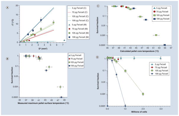
Aim: To compare the measured surface temperature of variable size ensembles of cells heated by intracellular magnetic fluid hyperthermia with heat diffusion model predictions.
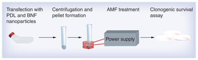
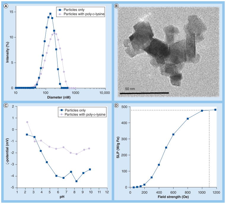
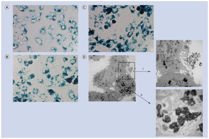
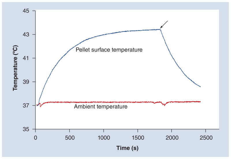
Materials & methods: Starch-coated Bionized NanoFerrite (Micromod Partikeltechnologie GmbH, Rostock, Germany) iron oxide magnetic nanoparticles were loaded into cultured DU145 prostate cancer cells. Cell pellets of variable size were treated with alternating magnetic fields. The surface temperature of the pellets was measured in situ and the associated cytotoxicity was determined by clonogenic survival assay.
Results & conclusion: For a given intracellular nanoparticle concentration, a critical minimum number of cells was required for cytotoxic hyperthermia. Above this threshold, cytotoxicity increased with increasing cell number. The measured surface temperatures were consistent with those predicted by a heat diffusion model that ignores intercellular thermal barriers. These results suggest a minimum tumor volume threshold of approximately 1 mm(3), below which nanoparticle-mediated heating is unlikely to be effective as the sole cytotoxic agent.
Figures
Similar articles
-
Characterization of intratumor magnetic nanoparticle distribution and heating in a rat model of metastatic spine disease.J Neurosurg Spine. 2014 Jun;20(6):740-50. doi: 10.3171/2014.2.SPINE13142. Epub 2014 Apr 4. J Neurosurg Spine. 2014. PMID: 24702509
-
Cell-Promoted Nanoparticle Aggregation Decreases Nanoparticle-Induced Hyperthermia under an Alternating Magnetic Field Independently of Nanoparticle Coating, Core Size, and Subcellular Localization.ACS Appl Mater Interfaces. 2019 Jan 9;11(1):340-355. doi: 10.1021/acsami.8b18451. Epub 2018 Dec 20. ACS Appl Mater Interfaces. 2019. PMID: 30525392
-
Real-time infrared thermography detection of magnetic nanoparticle hyperthermia in a murine model under a non-uniform field configuration.Int J Hyperthermia. 2013 Dec;29(8):752-67. doi: 10.3109/02656736.2013.839056. Epub 2013 Oct 18. Int J Hyperthermia. 2013. PMID: 24138472
-
Cancer hyperthermia using magnetic nanoparticles.Biotechnol J. 2011 Nov;6(11):1342-7. doi: 10.1002/biot.201100045. Epub 2011 Aug 26. Biotechnol J. 2011. PMID: 22069094 Review.
-
Magnetic nanoparticle hyperthermia for prostate cancer.Int J Hyperthermia. 2010;26(8):790-5. doi: 10.3109/02656731003745740. Epub 2010 Jul 23. Int J Hyperthermia. 2010. PMID: 20653418 Review.
Cited by
-
Micron-sized iron oxide particles for both MRI cell tracking and magnetic fluid hyperthermia treatment.Sci Rep. 2021 Feb 8;11(1):3286. doi: 10.1038/s41598-021-82095-6. Sci Rep. 2021. PMID: 33558583 Free PMC article.
-
Image-guided thermal therapy with a dual-contrast magnetic nanoparticle formulation: A feasibility study.Int J Hyperthermia. 2016 Aug;32(5):543-57. doi: 10.3109/02656736.2016.1159737. Epub 2016 May 5. Int J Hyperthermia. 2016. PMID: 27151045 Free PMC article.
-
Cancer therapy with iron oxide nanoparticles: Agents of thermal and immune therapies.Adv Drug Deliv Rev. 2020;163-164:65-83. doi: 10.1016/j.addr.2020.06.025. Epub 2020 Jun 27. Adv Drug Deliv Rev. 2020. PMID: 32603814 Free PMC article.
-
Magnetic Heating of Nanoparticles: The Importance of Particle Clustering to Achieve Therapeutic Temperatures.J Nanotechnol Eng Med. 2013 Feb;4(1):110071-1100714. doi: 10.1115/1.4024904. Epub 2013 Jul 16. J Nanotechnol Eng Med. 2013. PMID: 23919112 Free PMC article.
-
Magnetic nanoformulations for prostate cancer.Drug Discov Today. 2017 Aug;22(8):1233-1241. doi: 10.1016/j.drudis.2017.04.018. Epub 2017 May 16. Drug Discov Today. 2017. PMID: 28526660 Free PMC article. Review.
References
-
- Gordon RT, Hines JR, Gordon D. Intracellular hyperthermia. A biophysical approach to cancer treatment via intracellular temperature and biophysical alterations. Med Hypotheses. 1979;5(1):83–102. Relevant historical reference that first described the concept of intracellular hyperthermia as a potentially beneficial therapy. - PubMed
-
- Jordan A, Wust P, Scholz R, et al. Magnetic fluid hyperthermia (MFH) Scientific and Clinical Applications of Magnetic Carriers. 1997:569–595. Good historical review of intracellular nanoparticle hyperthermia.
-
- Jordan A, Schulz R, Wust P, et al. Endocytosis of dextran and silan-coated magnetite nanoparticles and the effect of intracellular hyperthermia on human mammary carcinoma cells in vitro. J Magn Magn Mater. 1999;194(1–3):185–196.
-
- Kita E, Oda T, Kayano T, et al. Ferromagnetic nanoparticles for magnetic hyperthermia and thermoablation therapy. J Phys D Appl Phys. 2010;43(47):474011.
Publication types
MeSH terms
Grants and funding
LinkOut - more resources
Full Text Sources
Other Literature Sources
Medical