. 2012 Nov 22:7:59.
doi: 10.1186/1750-1326-7-59.

Defective lysosomal proteolysis and axonal transport are early pathogenic events that worsen with age leading to increased APP metabolism and synaptic Abeta in transgenic APP/PS1 hippocampus

Affiliations

Defective lysosomal proteolysis and axonal transport are early pathogenic events that worsen with age leading to increased APP metabolism and synaptic Abeta in transgenic APP/PS1 hippocampus

Manuel Torres et al. Mol Neurodegener. .

Abstract

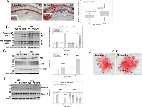
Background: Axonal pathology might constitute one of the earliest manifestations of Alzheimer disease. Axonal dystrophies were observed in Alzheimer's patients and transgenic models at early ages. These axonal dystrophies could reflect the disruption of axonal transport and the accumulation of multiple vesicles at local points. It has been also proposed that dystrophies might interfere with normal intracellular proteolysis. In this work, we have investigated the progression of the hippocampal pathology and the possible implication in Abeta production in young (6 months) and aged (18 months) PS1(M146L)/APP(751sl) transgenic mice.

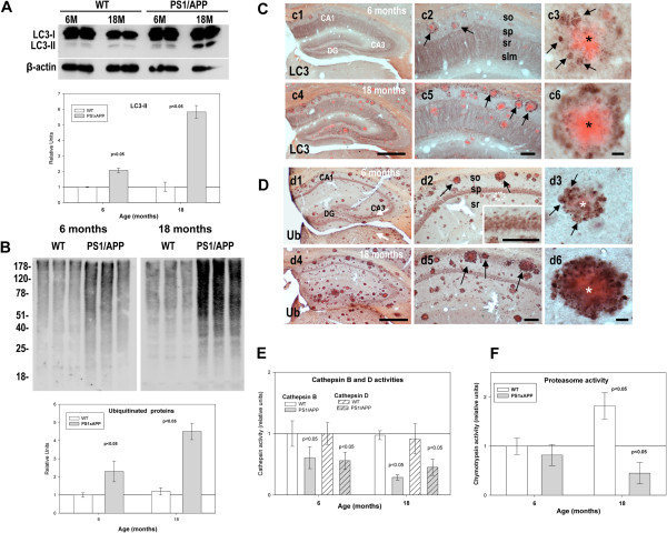
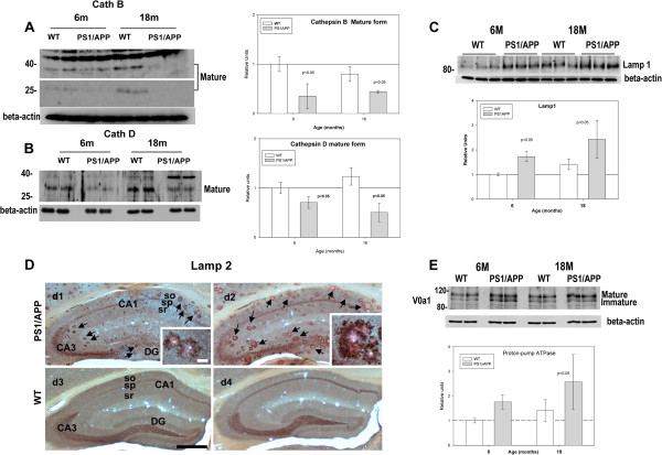
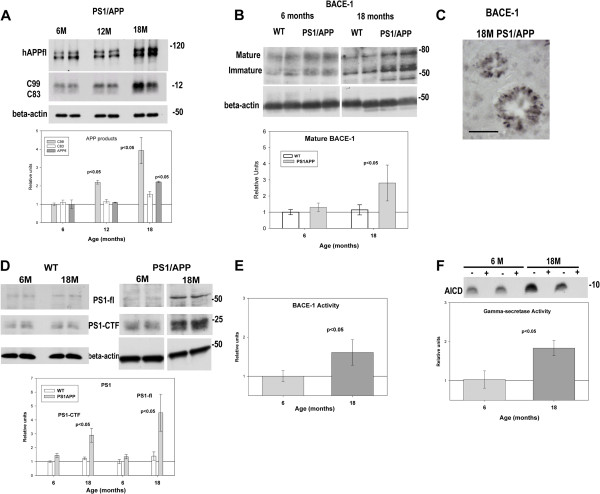
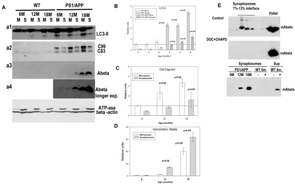
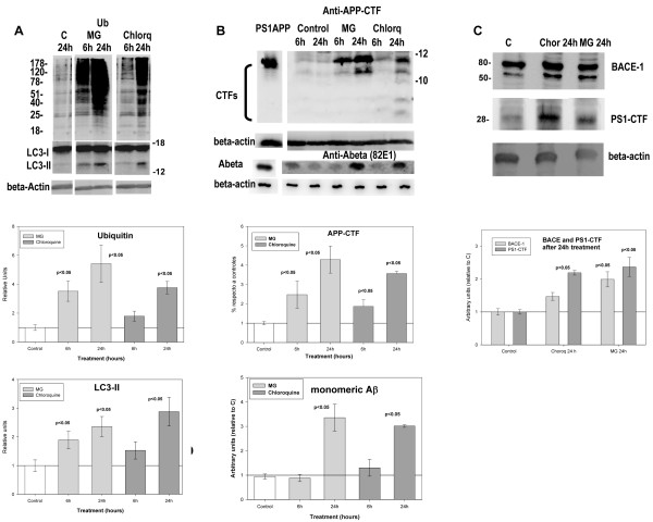
Results: Our data demonstrated the existence of a progressive, age-dependent, formation of axonal dystrophies, mainly located in contact with congophilic Abeta deposition, which exhibited tau and neurofilament hyperphosphorylation. This progressive pathology was paralleled with decreased expression of the motor proteins kinesin and dynein. Furthermore, we also observed an early decrease in the activity of cathepsins B and D, progressing to a deep inhibition of these lysosomal proteases at late ages. This lysosomal impairment could be responsible for the accumulation of LC3-II and ubiquitinated proteins within axonal dystrophies. We have also investigated the repercussion of these deficiencies on the APP metabolism. Our data demonstrated the existence of an increase in the amyloidogenic pathway, which was reflected by the accumulation of hAPPfl, C99 fragment, intracellular Abeta in parallel with an increase in BACE and gamma-secretase activities. In vitro experiments, using APPswe transfected N2a cells, demonstrated that any imbalance on the proteolytic systems reproduced the in vivo alterations in APP metabolism. Finally, our data also demonstrated that Abeta peptides were preferentially accumulated in isolated synaptosomes.

Conclusion: A progressive age-dependent cytoskeletal pathology along with a reduction of lysosomal and, in minor extent, proteasomal activity could be directly implicated in the progressive accumulation of APP derived fragments (and Abeta peptides) in parallel with the increase of BACE-1 and gamma-secretase activities. This retard in the APP metabolism seemed to be directly implicated in the synaptic Abeta accumulation and, in consequence, in the pathology progression between synaptically connected regions.


Figures

Figure 1
Figure 1
Aged-dependent increase of the hippocampal axonal pathology in PS1/APP mice.A) APP-immunolabeled CA1 sections with Congo red at 6 (left) and 18 (right) months of age showing the age-dependent increase in the dystrophic neurite pathology. Dystrophies were concentrated surrounding Abeta plaques (insets in a1 and a2 show higher magnifications). The quantitative analysis of plaque-associated dystrophies demonstrated a significant increase in aged mice. B) Representative western blots (n=5/age/group) for phosphorylated (upper panel) and non-phosphorylated (lower panel) neurofilament (SMI antibodies). Bands corresponding to high (H) and medium (M) chains were indicated. Quantitative analysis revealed an increase in the phosphorylated vs non-phosphorylated H and M neurofilaments in aged PS1/APP mice. No changes were observed in 6 month-old PS1/APP or WT mice. C) Tau phosphorylation was determined by western blots using AT8 (upper panel), AT100 (medium panel) and tau (lower panel) antibodies at 6 and 18 months of age (n=5/age/group). Graph showed the quantitative analysis of AT8 western blots. Due to the lack of immunoreactivity in WT and 6 months PS1/APP mice, the AT100 signal could not be quantified. A prominent increase in AT100 epitope (absent in other conditions) was observed in 18 month-old transgenic mice. D) AT8 immunostaining revealed that phospho-tau concentrated principally in dystrophies around congophilic plaques. E) Western blots and quantitative analysis (n=5/age/group) of kinesin (upper panel) and dynein (lower panel) expression. Both motor proteins decreased in PS1/APP mice. So, stratum oriens; sp, stratum pyramidale; sr, stratum radiatum. Scale bars: 200 μm (a1/a2), 10 μm (inset a1), 20 μm (inset a2), 20 μm (D).
Figure 2
Figure 2
Impairment of lysosomal and proteasomal protein-degradation in PS1/APP mice. A and B; Representative western blots and quantitative analysis (n=5 for each age and animal group) of the autophagic vesicle marker LC3 (A) and ubiquitinated proteins (B) performed using the same protein samples. An age-dependent accumulation of both LC3-II and ubiquitinated proteins were observed exclusively in PS1/APP samples. (C-D) Three different magnifications of LC3- (C) or ubiquitin- (D) immunolabeled hippocampal sections (counterstained with Congo red for Abeta plaques) of 6 and 18 months of age PS1/APP mice corroborated the age-dependent accumulation of both markers, associated with dystrophic neurites surrounding amyloid plaques (c1, c4, d1 and d4, panoramic views of immunostained hippocampus; c2, c5, d2 and d5, CA1 region, arrows indicate plaques covered by immunopositive dystrophies and inset in d2 shows immunolabeling in the pyramidal layer somata; c3, c6, d3 and d6, representative image of a congophilic plaque surrounded by dystrophic neurites, arrows point to positive dystrophies around a congophilic plaque indicated by an asterisk). Assays of lysosomal cathepsin B or D (E) and proteasomal chymotrypsin-like (F) enzymatic activities demonstrated the existence of a pronounced decrease in cathepsin B and D activities (since early ages in PS1/APP), whereas proteasome was affected only in aged tg mice. CA1, hippocampal subfield; DG, dentate gyrus; so, stratum oriens; sp, stratum pyramidale; sr, stratum radiatum; slm, stratum lacunoso-moleculare. Scale bars: 500 μm (c1/c4, d1/d4), 100 μm (c2/c5, d2/d5), 10 μm (c3/c5, d3/d5); 50 μm (d2, inset).
Figure 3
Figure 3
The mature forms of cathepsin B and D decreased whereas Lamp1, Lamp2 and V0a1 proton pump subunit accumulated in PS1/APP mice hippocampus. (A-B) Western blots and quantitative analysis (n=5 for each age and animal group) of cathepsin B (A) and D (B) showed the existence of a significant decrease in the mature forms of both proteins. This decrease was observed exclusively in PS1/APP transgenic mice at both ages tested (6 and 18 months). C-D) Same mice model accumulated Lamp1 (tested by western blot, C, n=3) or Lamp2 (tested by immunohistochemistry at 6 and 18 months of age). Lamp2 immunolabeled sections of 6 (d1) and 18 (d2) month-old PS1/APP mice counterstained with Congo red for fibrillar amyloid plaques revealed Lamp2 labeling associated to dystrophic neurites surrounding amyloid plaques (arrows) at both ages that increased with age. In WT mice (d3 and d4, for 6 and 18 months respectively) Lamp-2 immunostaining was associated to cell somata and neuropile. No dystrophies were detected in control samples. E) Representative western blot and quantitative analysis (n=5 for each age and animal group) of V0a1 proton pump subunit in samples from 6 and 18 month-old WT and PS1/APP mice. Both, the mature and immature forms of V0a1 seemed to be accumulated, although not significantly, in 6 and 18 months of age PS1/APP mice. CA1, hippocampal subfield; DG, dentate gyrus; so, stratum oriens; sp, stratum pyramidale; sr, stratum radiatum. Scale bars: 500 μm (D), 25 μm (insets in d1 and d2).
Figure 4
Figure 4
Increased amyloidogenic processing of APP in aged PS1/APP hippocampus.A) Representative western blots (n=4 for each age and animal group) for full-length APP (hAPPfl) (upper panel) and, APP-C terminal fragments (C99 and C83; middle panel) in 6, 12 and 18 month-old PS1/APP hippocampal samples. Quantitative analysis showed a significant increase in C99 from 6 to 18 months. APPfl also displayed a moderate (but significant) accumulation at 18 month-old. The amyloidogenic route was evaluated by testing the expression of BACE-1 (by western (B), n=5; and immunohistochermistry (C), n=3) and PS1 (n=6) (D) proteins. Dystrophic neurites surrounding plaques were positive for BACE-1 antibody. The enzymatic activities of BACE-1 (n=3) (E) and gamma-secretase (n=6) (F) were also assayed. Inset in panel F showed the accumulation of soluble AICD after 2 hours of incubation (at 37°C), in absence (minus) and in presence (plus) of 100 μM of the gamma-secretase inhibitor L-685-458, demonstrating that AICD production was dependent of the gamma-secretase activity. As shown, 18 month-old PS1/APP mice displayed an accumulation of both BACE-1 and PS1 (CTF fragment and full length) proteins paralleled by an increase in the corresponding enzymatic activities. Scale bar: 50 μm.
Figure 5
Figure 5
In vitro inhibition of either lysosomal and/or proteasomal activities induced the amyloidogenic APP processing in N2a-APPswe cultures.A) The inhibition of proteasomal/lysosomal (MG132) or lysosomal (Chloroquine) activities produced the accumulation of both ubiquitinated proteins (upper panel) and LC3-II autophagosomal marker (middle panel). Graphs showed the quantitative data of three different experiments using three different N2aAPPswe cultures. B) Using the same protein samples as in (A), we analyzed the effect of either inhibitor on the APP-CTF processing (upper western) and intracellular Abeta accumulation (lower western). As also shown quantitatively in graphs, either treatment produced the increase in the APP processing and the intracellular Abeta accumulation. C) In parallel, the expression of BACE-1 and PS1-ctf was also tested. As showed quantitatively, after 24h of treatment, both BACE-1 and PS1-cft were significantly accumulated in N2aAPPswe cells.
Figure 6
Figure 6
Monomeric Abeta was preferentially accumulated in isolated synaptosomal fraction, from aged PS1/APP mice, whereas APP-C99 fragment preferentially localized in microsomes.A) Microsomal and synaptosomal fractions were isolated from 6, 12 and 18 months WT and PS1/APP mice. Using these samples, we tested the distribution of LC3-II autophagosomal marker (a1), the APP-CTF fragments (a2) and the monomeric Abeta peptide (a3 and a4 for a longer exposure). ATP-synthase and b-actin were used as control of synapsomal integrity and protein loading. Quantitative analysis of western blots demonstrated the age-dependent accumulation of LC3-II (compared with the respective fraction in 6 month WT mice) (B) and C99 (as compared with 6-month-old PS1/APP microsomes) (C) in microsomal fractions. The monomeric Abeta was preferentially accumulated in the synaptosomal fractions (D). These experiments were repeated four times. E) Control experiments demonstrating the intrasynaptosomal origin of the monomeric Abeta. The tissue from a 12 month-old PS1/APP mice was divided, homogenized in absence (control) or in presence of 0.5% CHAPS + 0.5% DOC, and synaptosomes were isolated by Ficoll gradients. As shown, the disruption of cell membranes avoided the Abeta flotation on gradients (upper panels). On the other hand, tissue from 6 month-old WT mice were homogenized in absence (minus) of presence (plus) of synthetic Abeta42 and the synaptosomes were isolated. No Abeta were compartmentalized during homogenization.

Similar articles

Cited by

References

    1. Castellani RJ, Rolston RK, Smith MA. Alzheimer Disease. Dis Mon. 2010;56:484–546. doi: 10.1016/j.disamonth.2010.06.001. - DOI - PMC - PubMed
    1. Su JH, Cummings BJ, Cotman CW. Identification and distribution of axonal dystrophic neurites in Alzheimer's disease. Brain Res. 1993;625:228–237. doi: 10.1016/0006-8993(93)91063-X. - DOI - PubMed
    1. Nixon RA, Wegiel J, Kumar A, Yu WH, Peterhoff C, Cataldo A. et al.Extensive involvement of autophagy in Alzheimer disease: an immuno-electron microscopy study. J Neuropathol Exp Neurol. 2005;64:113–122. - PubMed
    1. Nixon RA. Autophagy, amyloidogenesis and Alzheimer disease. J Cell Sci. 2007;120:4081–4091. doi: 10.1242/jcs.019265. - DOI - PubMed
    1. Boland B, Kumar A, Lee S, Platt FM, Wegiel J, Yu WH. et al.Autophagy induction and autophagosome clearance in neurons: relationship to autophagic pathology in Alzheimer's disease. J Neurosci. 2008;28:6926–6937. doi: 10.1523/JNEUROSCI.0800-08.2008. - DOI - PMC - PubMed

Publication types

Substances

LinkOut - more resources