. 2013 Feb 15;304(4):C299-311.
doi: 10.1152/ajpcell.00302.2012. Epub 2012 Nov 21.

Mis-expression of the BK K(+) channel disrupts suprachiasmatic nucleus circuit rhythmicity and alters clock-controlled behavior

Affiliations

Mis-expression of the BK K(+) channel disrupts suprachiasmatic nucleus circuit rhythmicity and alters clock-controlled behavior

Jenna R Montgomery et al. Am J Physiol Cell Physiol. .

Abstract

In mammals, almost all aspects of circadian rhythmicity are attributed to activity in a discrete neural circuit of the hypothalamus, the suprachiasmatic nucleus (SCN). A 24-h rhythm in spontaneous firing is the fundamental neural intermediary to circadian behavior, but the ionic mechanisms that pattern circuit rhythmicity, and the integrated impact on behavior, are not well studied. Here, we demonstrate that daily modulation of a major component of the nighttime-phased suppressive K(+) current, encoded by the BK Ca(2+)-activated K(+) current channel (K(Ca)1.1 or Kcnma1), is a critical arbiter of circadian rhythmicity in the SCN circuit. Aberrant induction of BK current during the day in transgenic mice using a Per1 promoter (Tg-BK(R207Q)) reduced SCN firing or silenced neurons, decreasing the circadian amplitude of the ensemble circuit rhythm. Changes in cellular and circuit excitability in Tg-BK(R207Q) SCNs were correlated with elongated behavioral active periods and enhanced responses to phase-shifting stimuli. Unexpectedly, despite the severe reduction in circuit amplitude, circadian behavioral amplitudes in Tg-BK(R207Q) mice were relatively normal. These data demonstrate that downregulation of the BK current during the day is essential for the high amplitude neural activity pattern in the SCN that restricts locomotor activity to the appropriate phase and maintains the clock's robustness against perturbation. However, a residually rhythmic subset prevails over the ensemble circuit to drive the fundamental circadian behavioral rhythm.


Figures

Fig. 1.
Fig. 1.
BK Ca2+-activated K+ current expression and currents in suprachiasmatic nucleus (SCN) neurons. A: BK current as a function of voltage. Values are the peak paxilline-sensitive current at each voltage normalized to cell capacitance (means ± SE). BK currents are larger at night in SCN neurons. *Day vs. night across voltages: P = 10−7, factorial ANOVA, n = 6 [day, zeitgeber time (ZT)4–7] and 10 (night, ZT17–20); Bonferroni post hoc, P < 0.05 at +10, 30, and 50 mV. B: nighttime-phased K+ current was revealed by application of 0.5 mM 4-aminopyridine (4-AP). Values are mean peak currents at +30 mV ± SE. Nighttime K+ currents were 23% larger than daytime. After subtraction of the BK current with 10 μM paxilline (+Pax), the day-night difference in the residual current was reduced to 16%, although the difference was not significant (interaction, P = 0.52, two-way ANOVA). C: BK is expressed throughout the SCN and colocalizes with the major SCN neurotransmitters. Coronal sections from WT SCN at ZT6–8 incubated with α-BK (red, middle) and α-vasoactive intestinal peptide (VIP; i), α-green fluorescent protein (GFP) marking GAD67-expressing (GABA-ergic) cells (ii), and α-arginine vasopressin (AVP; iii) in green.
Fig. 2.
Fig. 2.
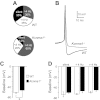
Kcnma1−/− SCN neurons are hyperactive at night. A: relative proportion of silent neurons (black), neurons firing spontaneously <4 Hz (gray), and >4 Hz (white) from wild type (WT) and Kcnma1−/− SCNs. P = 0.13, χ2 test; WT, n = 20; Kcnma1−/−, n = 25. B: Representative WT (black) and Kcnma1−/− (gray) spontaneous action potential waveforms. WT neuron fired at 5.9 Hz, Kcnma1−/− 6.2 Hz. C: average interspike baseline membrane potential (±SE) for all WT and Kcnma1−/− neurons. *P = 0.007, Mann-Whitney U-test. D: average interspike baseline membrane potential (±SE) separated into silent and active neurons (interaction of genotype across activity groups, P = 0.93, two-way ANOVA).
Fig. 3.
Fig. 3.
BK expression in Tg-BKR207Q SCNs. A-B: Tg-BKR207Q day (A) and night (B) SCN sections incubated with an α-BK antibody (red) and α-NF200 (green). BK is expressed throughout the SCN at both times. C: representative α-BK and α-tubulin (DM1α) Western blots from individual WT and homozygous Tg-BKR207Q SCNs harvested at 6-h time points. D: α-BK expression at each time point as a proportion of ZT20. WT BK expression over time (n = 4 SCNs at each time point) was different from Tg-BKR207Q (n = 3), P = 10−4, interaction effect with two-way ANOVA [ZT2, P = 0.02; ZT2 (cycle 2), P = 0.004, Bonferroni post hoc]. WT SCNs had a robust circadian difference in expression (P = 10−6, one-way ANOVA; Bonferroni post hoc between ZT2 and ZT20, P = 10−6), but the difference was less robust for Tg-BKR207Q across time points (P = 0.04, one-way ANOVA; Bonferroni post hoc between ZT2 and the ZT14, P = 0.11). E: real-time RT-PCR expression of Per2 and Bmal1 in WT and Tg-BKR207Q SCNs at ZT6 (day) and ZT19 (night), normalized to α-tubulin. Per2 and Bmal1 expression was both significantly different between day and night (*P = 0.02 and 0.01, respectively, time effect with two-way ANOVA) but not between genotypes (P = 0.29 and 0.42, genotype effect with two-way ANOVA). WT: n = 3 (day), 3 (night); Tg-BKR207Q: n = 2, 3.
Fig. 4.
Fig. 4.
BK currents in Tg-BKR207Q neurons. A: normalized current-voltage relationship between day and night BK currents from Tg-BKR207Q neurons. Interaction between time and voltage is no longer significant, P = 0.99, nsinteraction effect with factorial ANOVA, n = 14 (day, ZT4–7) and 11 (night, ZT17–20). B and C: normalized current-voltage relationship for BK currents from WT and Tg-BKR207Q during the day (B, *interaction between genotype and voltage, P = 10−10, factorial ANOVA; Bonferroni post hoc, P < 0.05 at +10 to +90 mV) and night (C, nsinteraction between genotype and voltage, P = 0.19, factorial ANOVA). Data re-plotted from Figs. 1C and 4A to facilitate cross comparisons. Simultaneous comparison of all conditions reveals a significant interaction between genotype, time, and voltage, P = 0.01, factorial ANOVA. D: integrated BK current from −50 to −10 mV (means ± SE) for WT (n = 12 day and n = 13 night) and Tg-BKR207Q (n = 19, 12) neurons. No significant interaction was found for genotype or time, P = 0.50, factorial ANOVA.
Fig. 5.
Fig. 5.
Excitability is altered in Tg-BKR207Q SCN neurons. A: relative proportion of WT and Tg-BKR207Q neurons that were silent (black), firing spontaneously <4 Hz (gray), and above 4 Hz (white). WT vs. Tg-BKR207Q: day, P = 0.03; night, P = 10−3; day vs. night: WT, p = 10−3, Tg-BKR207Q, P = 10−3; χ2 test. WT (day, night): n = 35, 36; Tg-BKR207Q: n = 34, 37. B: baseline membrane potential (means ± SE) for silent (S) and active (A) neurons. Silent neurons are more hyperpolarized than active (*A vs S, main effect, P = 10−3, factorial ANOVA), but the interaction among time, genotype, and activity group is not significant, P = 0.99, factorial ANOVA. C: firing rate for all neurons recorded, including silent cells (means ± SE). *Time effect for day vs. night, P = 10−3, two-way ANOVA. D: WT baseline membrane potentials. Silent cells are normally present in SCN neurons during the day. However, these cells maintain a hyperpolarized membrane potential that is similar to silent cells at night. E: Tg-BKR207Q baseline membrane potentials. Additional silent cells induced in Tg-BKR207Q SCNs also maintain a hyperpolarized membrane potential. FI: representative spontaneous action potentials from WT day (F; 5.1 Hz) and night (H; 2.4 Hz), and Tg-BKR207Q day (G; 3.2 Hz) and night (I; 2.7 Hz) neurons.
Fig. 6.
Fig. 6.
Circadian rhythms in WT and Tg-BKR207Q SCN circuits. A: representative coronal organotypic SCN slice on a 64-electrode probe. 3V, 3rd ventricle. Scale bar = 100 μm. B-C: Multi-unit spontaneous action potential activity on all 64 electrodes for 3 circadian cycles from a WT (B) and Tg-BKR207Q (C) slice. Shaded boxes are within the SCN, black: rhythmic, gray: arrhythmic. Y-axis is optimized for each recording to emphasize rhythmic vs. arrhythmic. D: Locations of arrhythmic recordings from WT (left side, ○, n = 27/229 recordings, 9 slices) and Tg-BKR207Q (right side, ●, n = 74/183 recordings, 7 slices) SCNs. E–G: Activity at a single electrode from a rhythmic WT (E), rhythmic Tg-BKR207Q (F), and arrhythmic Tg-BKR207Q (G) recording. H: average multiunit firing frequency during the peak and trough for rhythmic (R) recordings was different between WT and Tg-BKR207Q (*main effect of genotype, P = 10−3, two-way ANOVA). For both WT Tg-BKR207Q, there was a day vs. night difference in firing (main effect of time, P = 10−3, two-way ANOVA). For arrhythmic activity (AR), Tg-BKR207Q firing frequency was also reduced compared with WT (P = 0.007, t-test). WT, n = 9 slices, 202 recordings; Tg-BKR207Q, n = 7, 109. I and K: representative synchronized SCN circuit rhythm from WT (I) and Tg-BKR207Q (K) SCNs. Activity at each electrode is represented by a different color. J and L: representative χ2 periodograms from WT (J) and Tg-BKR207Q (L) SCNs. Arrow, circadian peak.
Fig. 7.
Fig. 7.
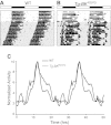
Circadian behavioral rhythms from WT and Tg-BKR207Q mice. A-B: Representative double-plotted running wheel actograms from a WT mouse (A) and Tg-BKR207Q (B) illustrating the elongation of alpha in Tg-BKR207Q mice. White and black bars denote 12:12 light-dark cycle. Shaded portion indicates constant darkness. C: average home cage activity from WT (n = 4) and Tg-BKR207Q (n = 3). Activity profiles are double-plotted and normalized to the peak (acrophase) activity level.
Fig. 8.
Fig. 8.
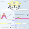
Summary schematizing the impact of diurnal expression of BK current on the SCN circuit and circadian behavior. Left: WT mice exhibit a robust circadian difference in SCN firing, correlated with BK expression levels. When BK currents are small, firing frequency is higher. When BK increases, firing is suppressed. Temporally delineated BK expression, low during the day and high at night, shapes high amplitude SCN activity. This circuit pattern is correlated with a restriction of locomotor activity during subjective night. Right: Tg-BKR207Q neurons do not exhibit a day-night difference in BK current. Daytime currents are as large as night, leading to suppression of daytime firing and loss of rhythmicity in almost half of the SCN. The consolidated circuit amplitude is low, potentially reducing inhibition of locomotor activity and leading to extension of behavioral activity beyond the WT boundaries. We propose this low-amplitude network is easier to phase-shift by light-driven input, leading to the observed larger phase delays and more rapid reentrainment compared with WT.

Similar articles

Cited by

References

    1. Abrahamson EE, Moore RY. Suprachiasmatic nucleus in the mouse: retinal innervation, intrinsic organization and efferent projections. Brain Res 916: 172–191, 2001 - PubMed
    1. Akiyama M, Kouzu Y, Takahashi S, Wakamatsu H, Moriya T, Maetani M, Watanabe S, Tei H, Sakaki Y, Shibata S. Inhibition of light- or glutamate-induced mPer1 expression represses the phase shifts into the mouse circadian locomotor and suprachiasmatic firing rhythms. J Neurosci 19: 1115–1121, 1999 - PMC - PubMed
    1. Albus H, Bonnefont X, Chaves I, Yasui A, Doczy J, van der Horst GT, Meijer JH. Cryptochrome-deficient mice lack circadian electrical activity in the suprachiasmatic nuclei. Curr Biol 12: 1130–1133, 2002 - PubMed
    1. Aton SJ, Colwell CS, Harmar AJ, Waschek J, Herzog ED. Vasoactive intestinal polypeptide mediates circadian rhythmicity and synchrony in mammalian clock neurons. Nat Neurosci 8: 476–483, 2005 - PMC - PubMed
    1. Aton SJ, Herzog ED. Come together right now: synchronization of rhythms in a mammalian circadian clock. Neuron 48: 531–534, 2005 - PMC - PubMed

Publication types

MeSH terms

Substances

LinkOut - more resources