

Impact of diabetes mellitus on bladder uroepithelial cells
- PMID: 23174855
- PMCID: PMC3543662
- DOI: 10.1152/ajpregu.00129.2012
Impact of diabetes mellitus on bladder uroepithelial cells
Abstract
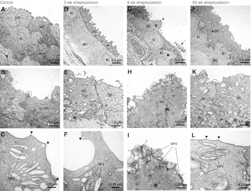
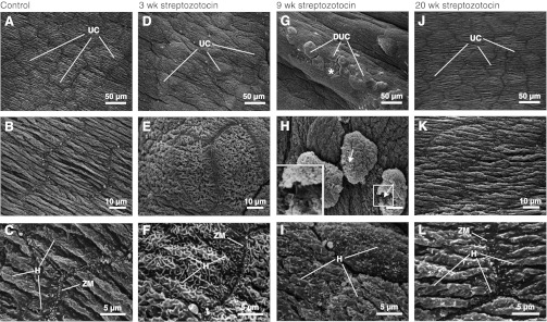
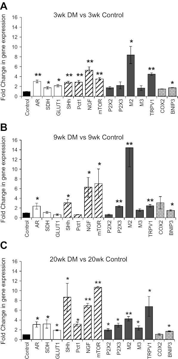
Diabetic bladder dysfunction (DBD), a prevalent complication of diabetes mellitus (DM), is characterized by a broad spectrum of symptoms including urinary urgency, frequency, and incontinence. As DBD is commonly diagnosed late, it is important to understand the chronic impact of DM on bladder tissues. While changes in bladder smooth muscle and innervation have been reported in diabetic patients, the impact of DM on the specialized epithelial lining of the urinary bladder, the urothelium (UT), is largely unknown. Quantitative polymerase chain reaction analysis and electron microscopy were used to evaluate UT gene expression and cell morphology 3, 9, and 20 wk following streptozotocin (STZ) induction of DM in female Sprague-Dawley rats compared with age-matched control tissue. Desquamation of superficial (umbrella) cells was noted at 9 wk DM, indicating a possible breach in barrier function. One causative factor may be metabolic burden due to chronic hyperglycemia, suggested by upregulation of the polyol pathway and glucose transport genes in DM UT. While superficial UT repopulation occurred by 20 wk DM, the phenotype was different, with significant upregulation of receptors associated with UT mechanosensation (transient receptor potential vanilloid subfamily member 1; TRPV1) and UT autocrine/paracrine signaling (acetylcholine receptors AChR-M2 and -M3, purinergic receptors P2X(2) and P2X(3)). Compromised barrier function and alterations in UT mechanosensitivity and cell signaling could contribute to bladder instability, hyperactivity, and altered bladder sensation by modulating activity of afferent nerve endings, which appose the urothelium. Our results show that DM impacts urothelial homeostasis and may contribute to the underlying mechanisms of DBD.
Figures
References
-
- Abdel-Aziz MT, Abdel-Kader MM, Rashad MM. Urinary catecholamines and their metabolites in diabetes. Acta Biol Med Ger 710: 1643–1650, 1975 - PubMed
-
- Andersson KE. Bladder activation: afferent mechanisms. Urology 59: 43–50, 2002 - PubMed
-
- Apodaca G. Modulation of membrane traffic by mechanical stimuli. Am J Physiol Renal Physiol 282: F179–F190, 2002 - PubMed
-
- Augustin R. The protein family of glucose transport facilitators: It's not only about glucose after all. IUBMB Life 62: 315–333, 2010 - PubMed
-
- Birder LA. Urothelial signaling. Hand Exp Pharmacol 202: 207–231, 2011 - PubMed
Publication types
MeSH terms
Substances
Grants and funding
LinkOut - more resources
Full Text Sources
Other Literature Sources
Medical
Research Materials