. 2013 Feb;87(3):1690-8.
doi: 10.1128/JVI.01855-12. Epub 2012 Nov 21.

Cleavage of interferon regulatory factor 7 by enterovirus 71 3C suppresses cellular responses

Affiliations

Cleavage of interferon regulatory factor 7 by enterovirus 71 3C suppresses cellular responses

Xiaobo Lei et al. J Virol. 2013 Feb.

Abstract

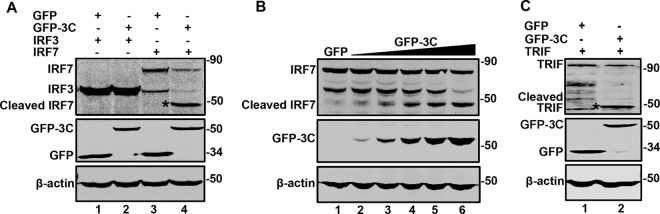
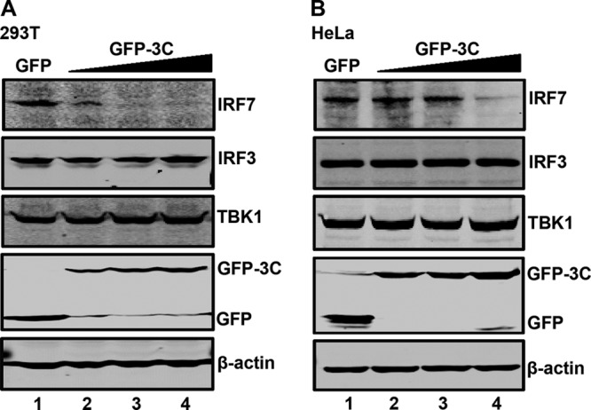
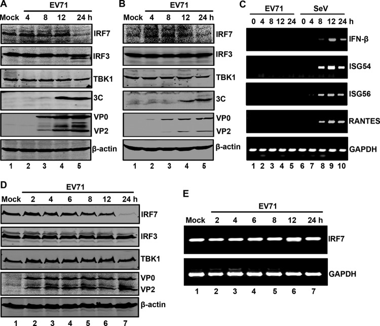
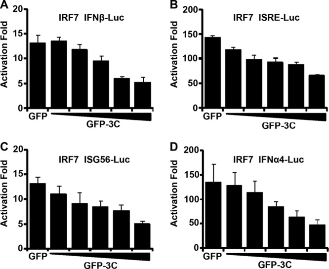
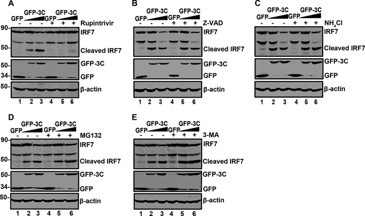
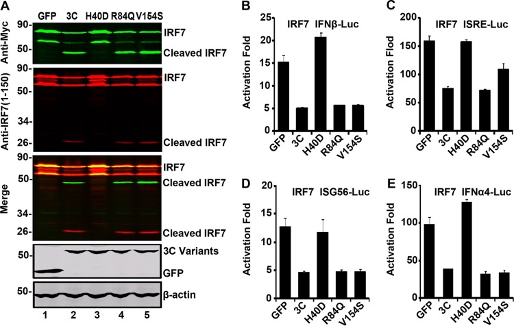
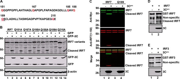
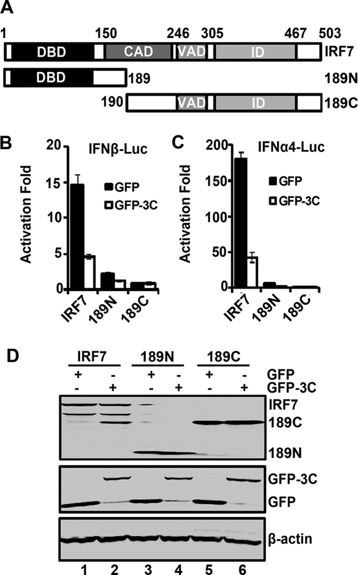
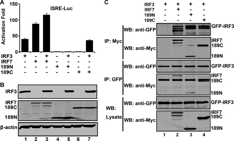
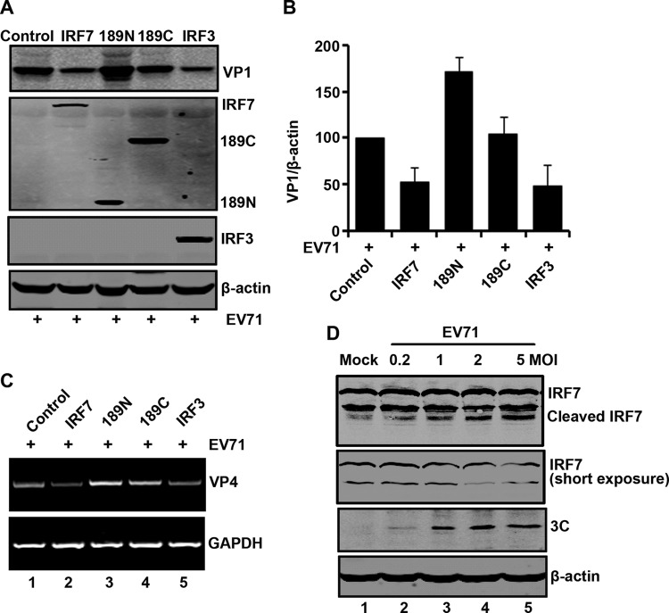
Enterovirus 71 (EV71) is a positive-stranded RNA virus which is capable of inhibiting innate immunity. Among virus-encoded proteins, the 3C protein compromises the type I interferon (IFN-I) response mediated by retinoid acid-inducible gene-I (RIG-I) or Toll-like receptor 3 that activates interferon regulatory 3 (IRF3) and IRF7. In the present study, we report that enterovirus 71 downregulates IRF7 through the 3C protein, which inhibits the function of IRF7. When expressed in mammalian cells, the 3C protein mediates cleavage of IRF7 rather than that of IRF3. This process is insensitive to inhibitors of caspase, proteasome, lysosome, and autophagy. H40D substitution in the 3C active site abolishes its activity, whereas R84Q or V154S substitution in the RNA binding motif has no effect. Furthermore, 3C-mediated cleavage occurs at the Q189-S190 junction within the constitutive activation domain of IRF7, resulting in two cleaved IRF7 fragments that are incapable of activating IFN expression. Ectopic expression of wild-type IRF7 limits EV71 replication. On the other hand, expression of the amino-terminal domain of IRF7 enhances EV71 infection, which correlates with its ability to interact with and inhibit IRF3. These results suggest that control of IRF7 by the 3C protein may represent a viral mechanism to escape cellular responses.


Figures

Fig 1
Fig 1
The 3C protease of EV71 cleaves IRF7 and TRIF. (A) 293T cells were transfected with plasmids encoding Myc-Flag-IRF3 (lanes 1 and 2) and Myc-Flag-IRF7 (lanes 3 and 4) or alone with GFP (lanes 1 and 3) or GFP-3C (lanes 2 and 4). At 24 h after transfection, cells were lysed and then cell lysates were analyzed by Western blotting with antibodies specific for Flag and GFP. β-Actin was used as a protein loading control. (B) 293T cells were transfected with Myc-Flag-IRF7 along with GFP (lanes 1) or increasing amounts of GFP-3C (lanes 2 to 6). The cells were then processed for Western blot analysis as described for panel A. (C) 293T cells were transfected with plasmids encoding Flag-TRIF (lanes 1 and 2) or alone with GFP (lanes 1) or GFP-3C (lanes 2). The cells were processed for Western blot analysis as described for panel A. Data shown are representative of three separate experiments.
Fig 2
Fig 2
The protease 3C mediates endogenous IRF7 cleavage. 293T (A) and HeLa (B) cells were transfected with the control GFP or increasing amounts of GFP-3C. At 24 h after transfection, cell lysates were subjected to Western blot analysis with antibodies against IRF7, IRF3, TBK1, GFP, and β-actin.
Fig 3
Fig 3
IRF7 is cleaved in EV71-infected cells. RD (A) and 293T (B) cells were mock infected or infected with EV71 at a multiplicity of infection (MOI) of 2. At the indicated time points, cell lysates were analyzed by Western blotting with antibodies for IRF7, IRF3, TBK1, EV71, EV71 3C, and β-actin. (C) 293T cells were mock infected or infected with EV71 (MOI = 2). At different time points after infection, total RNA extracted from cells were subjected to RT-PCR analysis for IFN-β, ISG54, ISG56, RANTES, and GAPDH mRNA. As a control, Sendai virus was used to infect cells. (D) THP1 cells were mock infected or infected with EV71 at an MOI of 10. At the indicated time points, cell lysates were analyzed by Western blotting with antibodies against IRF7, IRF3, TBK1, EV71, and β-actin. (E) THP1 cells were treated as described for panel D. Total RNAs extracted from cells were subjected to RT-PCR analysis for IRF7 and GAPDH mRNA.
Fig 4
Fig 4
EV71 3C inhibits promoter activation by IRF7. 293T cells were transfected with IRF7 and GFP-3C along with IFN-β-Luc (A), ISRE-Luc (B), ISG56-Luc (C), and IFN-α4-Luc (D). A plasmid expressing GFP or pRL-SV40 was used as a control. At 24 h after transfection, cell lysates were assayed for luciferase activities. Data are representative of three independent experiments with triplicate samples.
Fig 5
Fig 5
The effect of selected inhibitors on IRF7 cleavage. 293T cells were transfected with Myc-Flag-IRF7 along with GFP or GFP-3C. At 24 h after transfection, cells were incubated with the protease inhibitor rupintrivir (2 μM) (A) (31), caspase inhibitor Z-VAD (20 μM) (B) (6, 32), lysosome inhibitor NH4Cl (10 mM) (C) (33), proteasome inhibitor MG132 (0.1 μM) (D) (33), and autophagy inhibitor 3-MA (500 μM) (E) (34) for 24 h. Cell lysates were then processed for Western blot analysis.
Fig 6
Fig 6
(A) The effect of 3C variants on IRF7 cleavage. 293T cells were transfected with Myc-Flag-IRF7 (lanes 1 to 5), GFP, or GFP-3C variants as indicated. Cell lysates were subjected to Western blot analysis with antibodies as indicated using the LI-COR Odyssey dual-color system (LI-COR, Lincoln, NE). Antibodies recognizing Myc-Flag-IRF7 (Myc, C-terminal of IRF7, 800 nm, green; IRF7, 1 to 150 amino acids, 700 nm, red) were used. The merged images of the two channels were shown below (yellow). 3C or its variants were detected by using GFP antibody. β-Actin was included as a loading control. The effect of 3C variants on the IFN-β (B), ISRE (C), ISG56 (D), and IFN-α4 (E) promoter activation. 293T cells were transfected with plasmids encoding IRF7 and pIFN-β-Luc, pISRE-Luc, pISG56-Luc, or pIFN-α4-Luc, along with GFP or GFP-3C variants. pRL-SV40 was included as an internal control. At 24 h after transfection, cells were harvested to determine luciferase activities.
Fig 7
Fig 7
The Q189-S190 pair is the site of IRF7 cleavage. (A) Primary sequences of amino acids 151 to 216 within IRF7. In this region, glutamine was substituted with alanine. (B) IRF7 variant cleavage with substituting amino acids indicated above the lanes. 293T cells were transfected with wild-type IRF7 or IRF7 mutants along with GFP (lines 1, 3, 5, 7, 9, 11, and 13) or GFP-3C (lines 2, 4, 6, 8, 10, 12, and 14) as indicated. At 24 h after transfection, cell lysates were subjected to Western blot analysis with antibodies against Myc, GFP, and β-actin. (C) EV71 3C cleaves wild-type IRF7 but not the Q189A mutant in vitro. 293T cells were transfected with wild-type IRF7 or Q189A. At 24 h after transfection, cell lysates were incubated with the recombinant 3C protease. Samples were processed for Western blot analysis with antibodies using the LI-COR Odyssey dual-color system. Two antibodies that recognize Myc-IRF7 (Myc, C-terminal of IRF7, 800 nm, green; IRF7, 1 to 150 amino acids, 700 nm, red) were used. (D and E) EV71 3C cleaves recombinant IRF7 in vitro. The purified GST-IRF7 or GST-IRF3 was incubated with the 3C protease. Samples were analyzed by Western blotting using anti-GST antibody.
Fig 8
Fig 8
(A) Schematic diagrams of IRF7 deletion mutants. The following different domains are shown: the DNA binding domain (DBD), the constitutive activation domain (CAD), the virus-activated domain (VAD), the inhibitory domain (ID). 189N and 189C represent cleaved fragments of IRF7 by 3C. (B) The effect of 3C on IFN-β promoter activation induced by IRF7 and its mutants. 293T cells were transfected with IRF7, 189N, or 189C and GFP-3C along with IFN-β-Luc. GFP or pRL-SV40 was used as a control. At 24 h after transfection, cell lysates were assayed for luciferase activities. Data are representative of three independent experiments with triplicate samples. (C) The effect of 3C on IFN-α4 promoter activation induced by IRF7 and its mutants. 293T cells were transfected with IRF7, 189N, or 189C and GFP-3C along with IFN-α4-Luc. Cell lysates were detected as indicated in panel B. (D) Expression of IRF7 mutants. Cell lysates in panel B were detected by Western blotting with antibodies against Myc, GFP, and β-actin.
Fig 9
Fig 9
(A) The effects of IRF7 variants on the IRF3 activity. 293T cells were transfected with IRF7, 189N, 189C, or GFP-IRF3 along with ISRE-Luc as indicated. pRL-SV40 was used as a control. At 24 h after transfection, cell lysates were assayed for luciferase activities. Data are a representative of three independent experiments with triplicate samples. (B) Expression of IRF7 variants and IRF3. Cell lysates in panel A were detected by Western blotting with antibodies against Myc, GFP, and β-actin. (C) IRF7 variants interact with IRF3. 293T cells were transfected with plasmids encoding GFP-IRF3 (lanes 1, 2, 3, and 4), Myc-Flag-IRF7 (lane 2), Myc-Flag-189N (lane 3), and Myc-Flag-189C (lane 4). At 24 h after transfection, cell lysates were immunoprecipitated with anti-Myc or anti-GFP antibody. Immunoprecipitates and aliquots of cell lysates were then subjected to Western blot analysis.
Fig 10
Fig 10
(A) IRF7 inhibits EV71 replication. RD cells were transfected with IRF7 variants and IRF3 as indicated. At 24 h after transfection, cells were treated with EV71. After 24 h, cells were harvested and resolved with 12% SDS-PAGE. Western blot analysis for VP1, Myc, or β-actin was conducted. (B) Densitometry analysis. VP1 protein bands from three independent experiments in panel A were quantified and normalized to β-actin by using the Odyssey image software. (C) The effects of IRF7 on viral RNA synthesis. RD cells were transfected with IRF7, 189N, 189C, or IRF3 for 24 h. Cells were infected with EV71, and at 24 h postinfection, the total RNA extracted from the cells were analyzed for the expression of EV71 VP4 RNA. GAPDH was used as an internal control. (D) IRF7 is cleaved in EV71-infected cells. 293T cells were transfected with IRF7 for 24 h. Cells were then mock infected or infected with an increasing dose of EV71. At 24 h after infection, cell lysates were analyzed by Western blotting using antibodies against Myc, EV71, and β-actin.

References

    1. McMinn PC. 2002. An overview of the evolution of enterovirus 71 and its clinical and public health significance. FEMS Microbiol. Rev. 26:91–107 - PubMed
    1. Shih SR, Chiang C, Chen TC, Wu CN, Hsu JT, Lee JC, Hwang MJ, Li ML, Chen GW, Ho MS. 2004. Mutations at KFRDI and VGK domains of enterovirus 71 3C protease affect its RNA binding and proteolytic activities. J. Biomed. Sci. 11:239–248 - PubMed
    1. Sim AC, Luhur A, Tan TM, Chow VT, Poh CL. 2005. RNA interference against enterovirus 71 infection. Virology 341:72–79 - PubMed
    1. Li ML, Hsu TA, Chen TC, Chang SC, Lee JC, Chen CC, Stollar V, Shih SR. 2002. The 3C protease activity of enterovirus 71 induces human neural cell apoptosis. Virology 293:386–395 - PubMed
    1. Weng KF, Li ML, Huang CT, Shih SR. 2009. Enterovirus 71 3C protease cleaves a novel target CstF-64 and inhibits cellular polyadenylation. PLoS Pathog. 5:e1000593 doi:10.1371/journal.ppat.1000593 - DOI - PMC - PubMed

Publication types

LinkOut - more resources