. 2013 Feb;87(3):1733-49.
doi: 10.1128/JVI.02958-12. Epub 2012 Nov 21.

Reactive oxygen species are induced by Kaposi's sarcoma-associated herpesvirus early during primary infection of endothelial cells to promote virus entry

Affiliations

Reactive oxygen species are induced by Kaposi's sarcoma-associated herpesvirus early during primary infection of endothelial cells to promote virus entry

Virginie Bottero et al. J Virol. 2013 Feb.

Abstract

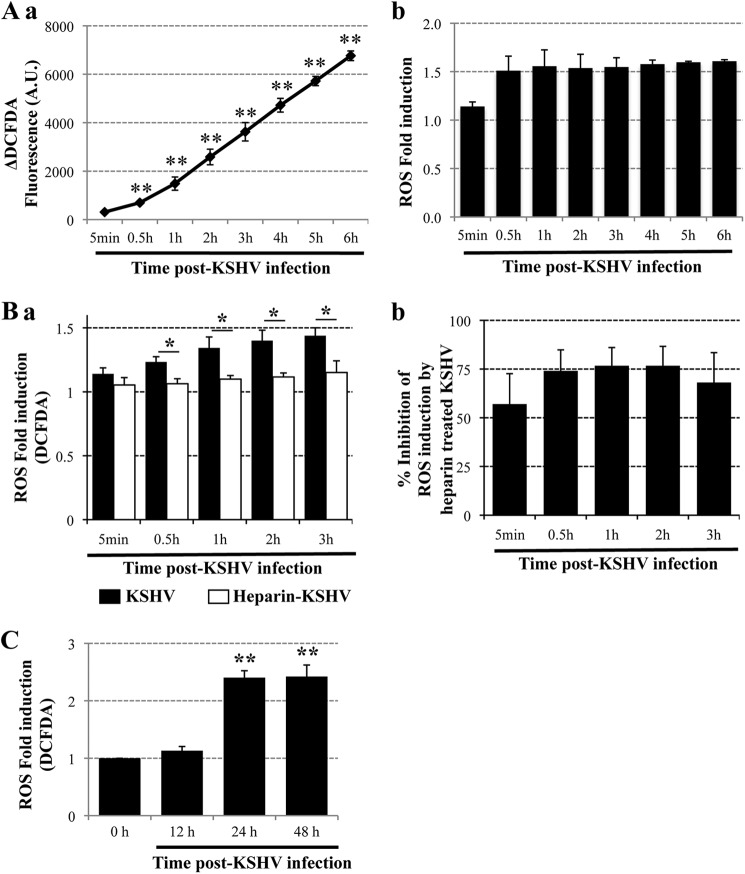
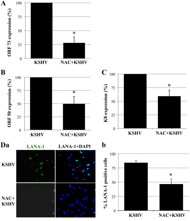
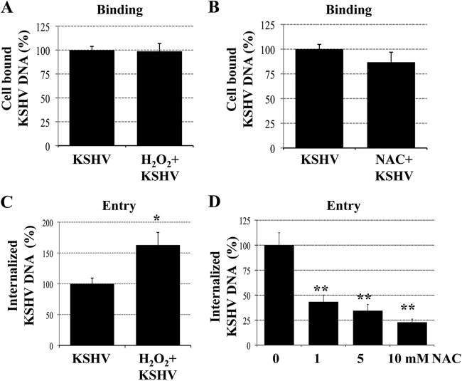
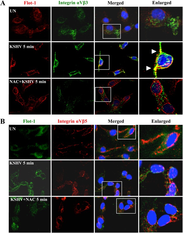
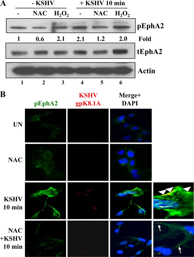
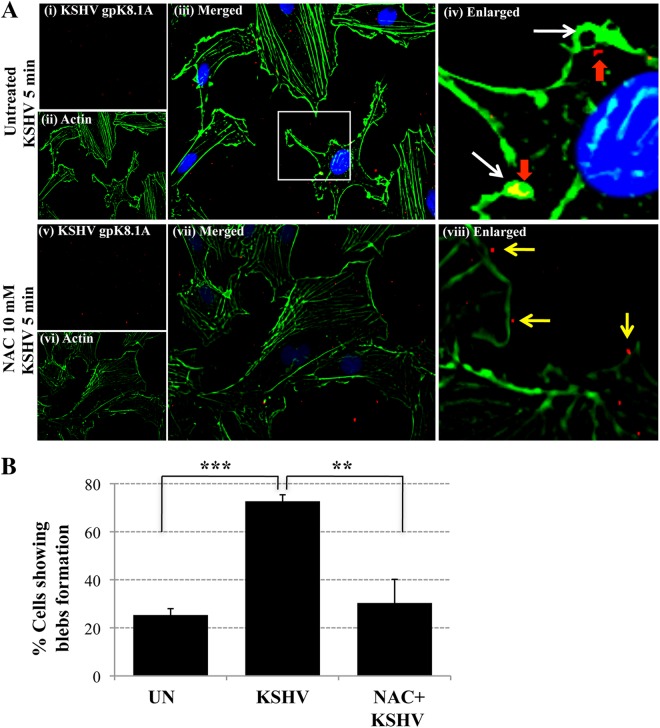
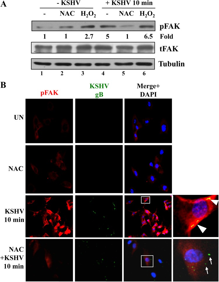
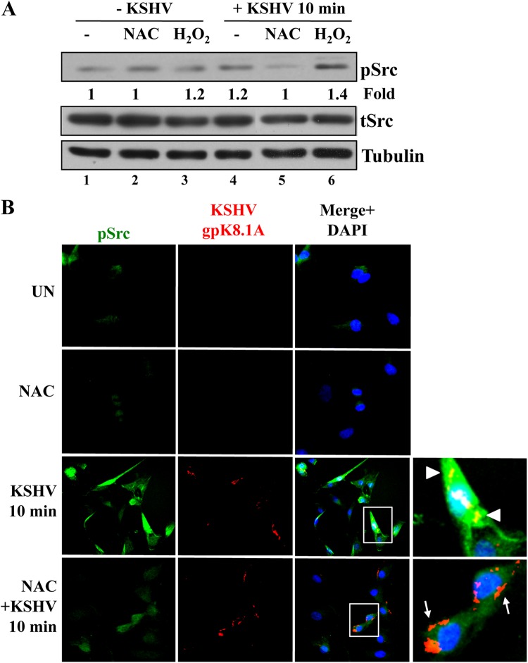
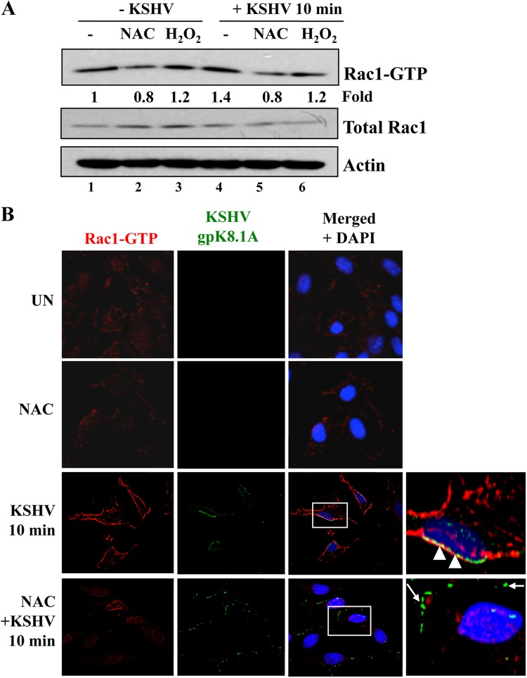
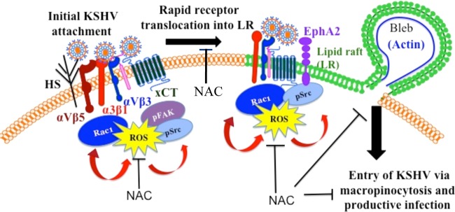
The entry of Kaposi's sarcoma-associated herpesvirus (KSHV) into human dermal microvascular endothelial cells (HMVEC-d), natural in vivo target cells, via macropinocytosis is initiated through a multistep process involving the binding of KSHV envelope glycoproteins with cell surface α3β1, αVβ3, and αVβ5 integrin molecules and tyrosine kinase ephrin-A2 receptor, followed by the activation of preexisting integrin-associated signaling molecules such as focal adhesion kinase (FAK), Src, c-Cbl, phosphoinositide 3-kinase (PI-3K), and Rho-GTPases. Many viruses, including KSHV, utilize cellular reactive oxygen species (ROS) for viral genomic replication and survival within host cells; however, the role of ROS in early events of viral entry and the induction of signaling has not been elucidated. Here we show that KSHV induced ROS production very early during the infection of HMVEC-d cells and that ROS production was sustained over the observation period (24 h postinfection). ROS induction was dependent on the binding of KSHV to the target cells, since pretreatment of the virus with heparin abolished ROS induction. Pretreatment of HMVEC-d cells with the antioxidant N-acetylcysteine (NAC) significantly inhibited KSHV entry, and consequently gene expression, without affecting virus binding. In contrast, H(2)O(2) treatment increased the levels of KSHV entry and infection. In addition, NAC inhibited KSHV infection-induced translocation of αVβ3 integrin into lipid rafts, actin-dependent membrane perturbations, such as blebs, observed during macropinocytosis, and activation of the signal molecules ephrin-A2 receptor, FAK, Src, and Rac1. In contrast, H(2)O(2) treatment increased the activation of ephrin-A2, FAK, Src, and Rac1. These studies demonstrate that KSHV infection induces ROS very early during infection to amplify the signaling pathways necessary for its efficient entry into HMVEC-d cells via macropinocytosis.


Figures

Fig 1
Fig 1
(A) ROS production upon primary infection of endothelial cells with KSHV. Confluent HMVEC-d cells in 12-well plates were serum starved for 2 h, incubated with EBM-2 containing 10 μM CM-H2DCFDA for 1 h at 37°C, and either mock infected or infected with KSHV (40 DNA copies/cell). Fluorescence was measured with a Synergy HT microplate reader (BioTek Instruments) using a 485/20 excitation and 528/20 emission filter pair and a PMT sensitivity setting of 55. Each reading was done in triplicate, and the data are means for three independent experiments ± standard deviations. Statistical analysis was conducted using a two-tailed Student test. **, P < 0.01. (a) ΔDCFDA fluorescence between infected and uninfected cells. A.U., arbitrary units. (b) Fold induction of ROS production in infected cells relative to uninfected cells. (B) Binding of KSHV to endothelial cells induces ROS production. HMVEC-d cells were starved for 2 h, loaded with CM-H2DCFDA (10 μM) for 1 h, and infected either with untreated KSHV (40 DNA copies/cell) or with KSHV preincubated with 100 μg/ml heparin for 1 h at 37°C (40 DNA copies/cell). The respective control cells were mock infected either with medium alone or with medium containing 100 μg/ml heparin. (a) Fluorescence was measured at the indicated time points, and ROS production is expressed as fold induction relative to the respective controls. Each reading was done in triplicate, and the data are means for two independent experiments ± standard deviations. Statistical analysis was performed using a two-tailed Student test. *, P < 0.05. (b) The inhibition of ROS production in cells infected with heparin-treated virus was compared with ROS production in cells infected with untreated KSHV, and the percentage of inhibition is shown. (C) Elevated ROS production during KSHV latency in endothelial cells. Serum-starved (2 h) HMVEC-d cells were either mock infected or infected with KSHV (40 DNA copies/cell); the virus was removed; the cells were loaded with CM-H2DCFDA for 1 h at 12 h, 24 h, and 48 h p.i.; and fluorescence was measured. **, P < 0.01.
Fig 2
Fig 2
ROS participate in primary KSHV infection of HMVEC-d cells. (A, B, and C) ROS induction is necessary for the expression of KSHV genes. Serum-starved (2 h) HMVEC-d cells were either mock treated or pretreated with NAC (10 mM) for 2 h, after which they were infected with KSHV (20 DNA copies/cells) for 2 h. Then the virus was removed, and the cells were mock treated or treated with NAC (1 mM) for 24 h. RNA was extracted with TRIzol reagent (Invitrogen) and was analyzed by real-time RT-PCR using latency-associated ORF 73 (A) or lytic-cycle-associated ORF 50 (B) or K8 (C) gene primers. Each PCR was performed in triplicate, and the data are means for two independent experiments ± standard deviations. (D) ROS induction is necessary for LANA-1 expression. HMVEC-d cells in chamber slides were either mock treated or pretreated with NAC (10 mM) for 2 h and were then infected with KSHV (20 DNA copies/cells) for 2 h. Unbound virus was removed by washing, and cells were either mock treated or treated with NAC (1 mM) for 48 h. Cells were fixed and processed for an IFA using a rabbit anti-LANA-1 antibody. (a) Representative IFA images. (b) LANA-1-positive cells from four different independent fields, each containing at least 15 cells, were counted to calculate the percentage of LANA-1-positive cells. Statistical analysis was carried out using a two-tailed Student test. *, P < 0.05.
Fig 3
Fig 3
ROS induction during primary infection of endothelial cells promotes KSHV entry. (A and B) ROS have no effect on KSHV binding. HMVEC-d cells were infected with KSHV (20 DNA copies/cell) for 1 h at 4°C in the presence of H2O2 (100 μM) (A) or were treated with NAC (10 mM) for 2 h prior to infection (B). Then the cells were washed; total DNA was isolated; and KSHV binding was determined by real-time DNA PCR for the ORF 73 gene. (C and D) ROS are involved in KSHV entry. HMVEC-d cells were infected with KSHV (20 DNA copies/cell) for 1 h at 37°C in the presence of H2O2 (100 μM) (C) or were treated with NAC (1, 5, and 10 mM) for 2 h prior to infection (D). After washing, cells were treated with 0.25% trypsin-EDTA for 5 min at 37°C to remove bound but noninternalized virus and were then washed; total DNA was isolated; and KSHV entry was determined by real-time DNA PCR for the ORF 73 gene. Each PCR was carried out in triplicate, and the data are means for three independent experiments ± standard errors of the means. Statistical analysis was carried out using a two-tailed Student test. *, P < 0.05; **, P < 0.01.
Fig 4
Fig 4
ROS induced by primary infection with KSHV participate in the translocation of integrin αVβ3 into lipid rafts. HMVEC-d cells were starved for 2 h, pretreated with NAC (10 mM) for 2 h, infected with KSHV (20 DNA copies/cell) for 5 min at 37°C, and then washed. The cells were then processed for IFA using the following antibodies: anti-flotillin-1 (with an anti-goat secondary antibody conjugated to Alexa Fluor 584 [A] or Alexa Fluor 488 [B]) and anti-integrin αVβ3 (with an Alexa Fluor 488-conjugated anti-mouse secondary antibody) (A) or anti-integrin αVβ5 (with an Alexa Fluor 584-conjugated anti-mouse secondary antibody) (B). Boxed areas are enlarged. Arrowheads indicate the colocalization of integrin αVβ3 with lipid rafts. Magnification, ×40.
Fig 5
Fig 5
ROS participate in EphA2 activation induced by KSHV infection. HMVEC-d cells were serum starved for 5 h. Uninfected serum-starved cells were treated either with 5 mM NAC for 2 h or with 100 μM H2O2 for 10 min. For KSHV infection, either serum-starved cells were pretreated with 5 mM NAC for 2 h or 100 μM H2O2 was added simultaneously with KSHV. Cells were infected with KSHV (20 DNA copies/cell) for 10 min at 37°C, washed, and processed for Western blot analysis (A) or IFA (B) using the indicated antibodies. (A) pEphA2 induction was calculated by assigning the value of 1 to levels in uninfected cells. tEphA2, total EphA2. (B) Arrowheads indicate colocalization of KSHV (gpK8.1A) with pEphA2, whereas arrows indicate the viral particle outside the cells, not colocalized with pEphA2. UN, uninfected cells. Magnification, ×40.
Fig 6
Fig 6
The antioxidant NAC blocks the actin reorganization and membrane bleb formation observed during KSHV entry. (A) HMVEC-d cells were starved for 2 h, pretreated with NAC (10 mM) for 2 h, infected with KSHV (20 DNA copies/cell) for 5 min at 37°C, washed, and processed for IFA using phalloidin (Alexa Fluor 488 conjugate) and an anti-KSHV gpK8.1A antibody (with an Alexa Fluor 594-conjugated anti-mouse secondary antibody). A representative image is shown. Boxed area is enlarged. White arrows indicate bleb formations. Red arrows indicate the colocalization of blebs and internalized viral particles. Yellow arrows indicate viral particles outside the cells. Magnification, ×40. (B) The percentage of cells bearing membrane projections was determined by counting such cells in a field containing at least 10 cells. Three independent fields were chosen for uninfected (UN) cells, KSHV-infected cells, and KSHV-infected cells pretreated with NAC. Statistical analysis was performed using a two-tailed Student test. **, P < 0.01; ***, P < 0.005.
Fig 7
Fig 7
ROS participate in KSHV-induced FAK activation. HMVEC-d cells were starved for 5 h. Either cells were treated with 5 mM NAC for 2 h prior to KSHV infection or 100 μM H2O2 was added simultaneously with KSHV. Cells were infected with KSHV (20 DNA copies/cell) for 10 min at 37°C, washed, and processed for Western blot analysis (A) or IFA (B) using the antibodies indicated. (A) pFAK induction was calculated by assigning the value of 1 to levels in uninfected cells. Tubulin was used as a loading control. (B) Boxed areas are enlarged. Arrowheads indicate colocalization of KSHV (gB) with pFAK. Arrows indicate viral particles outside the cells, not colocalized with pFAK. Magnification, ×40.
Fig 8
Fig 8
ROS participate in KSHV-induced Src activation. HMVEC-d cells were starved for 5 h. Either cells were treated with 5 mM NAC for 2 h prior to KSHV infection or 100 μM H2O2 was added simultaneously with KSHV. Cells were infected with KSHV (20 DNA copies/cell) for 10 min at 37°C, washed, and processed for Western blot analysis (A) or IFA (B) using the antibodies indicated. (A) pSrc induction was calculated by assigning the value of 1 to levels in uninfected cells. Tubulin was used as a loading control. (B) Boxed areas are enlarged. Arrowheads indicate colocalization of KSHV (gpK8.1A) with pSrc. Arrows indicate viral particles outside the cells, not colocalized with pSrc. Magnification, ×40.
Fig 9
Fig 9
ROS participate in Rac1 activation induced by KSHV. HMVEC-d cells were starved for 5 h. Either cells were treated with 5 mM NAC for 2 h prior to KSHV infection or 100 μM H2O2 was added simultaneously with KSHV. Cells were infected with KSHV (20 DNA copies/cell) for 10 min at 37°C, washed, and processed for a pulldown assay (A) or IFA (B) using the antibodies indicated. (A) Rac1 induction was calculated by assigning the value of 1 to levels in uninfected cells. (B) Boxed areas are enlarged. Arrowheads indicate colocalization of KSHV (gpK8.1A) with Rac1-GTP. Arrows indicate viral particles outside the cells, not colocalized with Rac1-GTP. Magnification, ×40.
Fig 10
Fig 10
Schematic depicting the induction of ROS by KSHV early during primary infection of endothelial cells to promote virus entry. KSHV initially binds and interacts with heparan sulfate, integrins (α3β1, αVβ3, αVβ5), and x-CT in non-LR regions of HMVEC-d cells and is then rapidly translocated, along with selective integrins (α3β1, αVβ3) and x-CT receptors, into lipid rafts. In LRs, the receptor EphA2 interacts with KSHV and integrins and is phosphorylated. KSHV entry is initiated by the induction of signaling pathways such as FAK, Src, and Rac1 and the concurrent formation of macropinocytic blebs. The studies presented here demonstrate increased ROS production very early during KSHV primary infection. Pretreatment with the antioxidant NAC significantly inhibited KSHV entry, and consequently gene expression, without affecting virus binding. The translocation of KSHV-integrins into LRs, actin-dependent membrane bleb formation, and the activation of the signal molecules ephrin-A2, FAK, Src, and Rac1 were inhibited by NAC. In contrast, H2O2 treatment increased KSHV entry and the phosphorylation of ephrin-A2, FAK, and Src. These studies demonstrate that KSHV infection induces ROS very early during infection to amplify the signaling pathways that are necessary for the efficient entry of viral particles into HMVEC-d cells via macropinocytosis.

Similar articles

Cited by

References

    1. Cesarman E, Chang Y, Moore PS, Said JW, Knowles DM. 1995. Kaposi's sarcoma-associated herpesvirus-like DNA sequences in AIDS-related body-cavity-based lymphomas. N. Engl. J. Med. 332:1186–1191 - PubMed
    1. Ganem D. 2007. Kaposi‘s sarcoma-associated herpesvirus, p 2847–2888 In Knipe DM, Howley PM, Griffin DE, Lamb RA, Martin MA, Roizman B, Straus SE. (ed), Fields virology, 5th ed, vol 2 Lippincott Williams & Wilkins, Philadelphia, PA
    1. Krishnan HH, Naranatt PP, Smith MS, Zeng L, Bloomer C, Chandran B. 2004. Concurrent expression of latent and a limited number of lytic genes with immune modulation and antiapoptotic function by Kaposi's sarcoma-associated herpesvirus early during infection of primary endothelial and fibroblast cells and subsequent decline of lytic gene expression. J. Virol. 78:3601–3620 - PMC - PubMed
    1. Li X, Feng J, Sun R. 2011. Oxidative stress induces reactivation of Kaposi's sarcoma-associated herpesvirus and death of primary effusion lymphoma cells. J. Virol. 85:715–724 - PMC - PubMed
    1. Ye F, Gao SJ. 2011. A novel role of hydrogen peroxide in Kaposi sarcoma-associated herpesvirus reactivation. Cell Cycle 10:3237–3238 - PMC - PubMed

Publication types

Substances