. 2013 Jan 1;140(1):56-65.
doi: 10.1242/dev.086413. Epub 2012 Nov 22.

Hedgehog is required for CySC self-renewal but does not contribute to the GSC niche in the Drosophila testis

Affiliations

Hedgehog is required for CySC self-renewal but does not contribute to the GSC niche in the Drosophila testis

Marc Amoyel et al. Development. .

Abstract

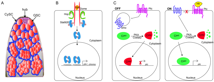
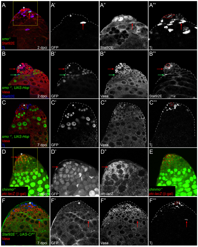
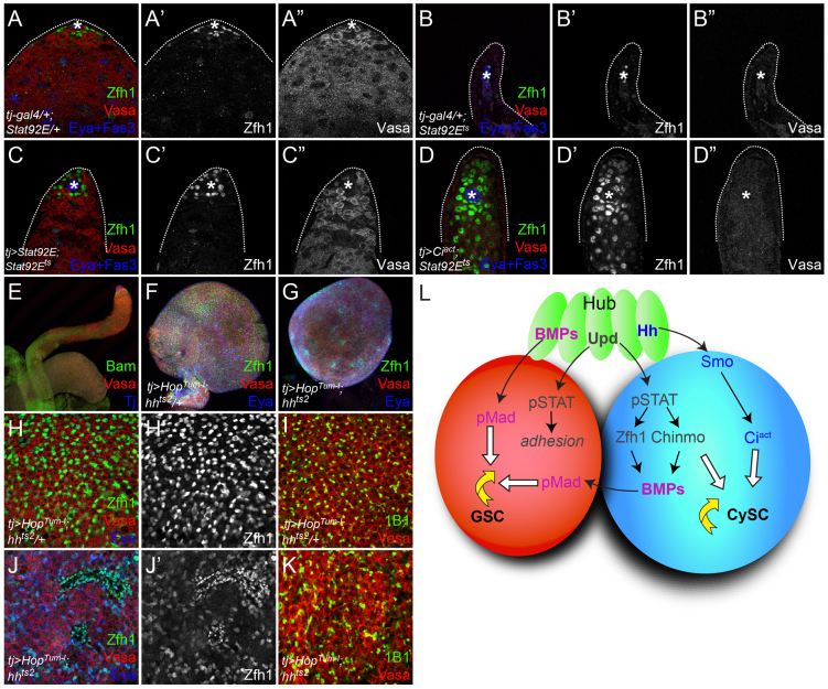
The Drosophila testis harbors two types of stem cells: germ line stem cells (GSCs) and cyst stem cells (CySCs). Both stem cell types share a physical niche called the hub, located at the apical tip of the testis. The niche produces the JAK/STAT ligand Unpaired (Upd) and BMPs to maintain CySCs and GSCs, respectively. However, GSCs also require BMPs produced by CySCs, and as such CySCs are part of the niche for GSCs. Here we describe a role for another secreted ligand, Hedgehog (Hh), produced by niche cells, in the self-renewal of CySCs. Hh signaling cell-autonomously regulates CySC number and maintenance. The Hh and JAK/STAT pathways act independently and non-redundantly in CySC self-renewal. Finally, Hh signaling does not contribute to the niche function of CySCs, as Hh-sustained CySCs are unable to maintain GSCs in the absence of Stat92E. Therefore, the extended niche function of CySCs is solely attributable to JAK/STAT pathway function.


Figures

Fig. 1.
Fig. 1.
The JAK/STAT and Hh pathways. (A) The Drosophila testis. See text for details. (B) JAK/STAT signaling in Drosophila. Binding of the secreted ligand Unpaired (Upd) to its receptor Domeless (Dome) causes phosphorylation and activation of the Janus kinase (JAK) Hopscotch (Hop). Hop in turns phosphorylates the transcription factor Stat92E, which dimerizes and enters the nucleus to promote the transcription of target genes, including zfh1 and chinmo. (C) The Hh pathway in the absence (‘OFF’) or presence (‘ON’) of ligand. When Hh is not present, Patched (Ptc) inhibits Smoothened (Smo), rendering it inactive. In the absence of Smo activity, the transcription factor Cubitus interruptus (Ci) is cleaved from a full-length transcriptional activator form (Ciact) to a repressor (Cirep) by a complex that includes the Protein kinase A (PKA) enzyme and scaffolding proteins such as Costal2. Hh ligand (‘ON’) prevents Ptc activity, allowing Smo to inhibit Ci cleavage, meaning that Ciact enters the nucleus to activate transcription of target genes, such as ptc. GSC, germ line stem cell; CySC, cyst stem cell; Y, tyrosine residues.
Fig. 2.
Fig. 2.
Expression of Hh pathway components at the testis niche. (A,A′) Expression of hh-lacZ is seen in the hub (arrow in A′). (B,B′) Hh protein is also found at high levels in and around the hub (arrow in B′). (C-D″′) tj-gal4>UAS-CD8::GFP was used to label somatic cell membranes (single channel in C′,D′). CySCs co-express Ptc (red; single channel in C″) and Zfh1 (blue; single channel in C″′) adjacent to the hub. Ci (red; single channel in D″) is expressed in the cyst lineage, including Zfh1-positive CySCs. Asterisks indicate the hub, which is outlined (red dotted lines) in C″′,D″′.
Fig. 3.
Fig. 3.
Hh signaling is required for CySC self-renewal. (A-F′) MARCM clonal analysis showing control (A,D) or smo mutant clones (B,C,E,F). (A-C″′) Control FRT40A CySC clones at 2 dpci can be recovered, as assessed by Zfh1 expression (blue) and lack of Eya (red) (A). smo mutant CySC clones can be recovered at 2 dpci (B) and express Zfh1, whereas other smo mutant clones initiate normal differentiation and begin to express Eya (C). Single channel for GFP is shown in A′-C′, for Eya in A″-C″ and for Zfh1 in A″′C″′. (D-D″′) Control FRT40A MARCM clone (green) at 7 dpci showing a marked CySC (red arrow) and GSC (yellow arrowhead) adjacent to the hub. (E-F′) smo mutant MARCM clone at 7 dpci showing a labeled GSC (yellow arrowhead) but no CySCs (E). The green arrow points to a marked differentiated cyst cell (E). smo mutant CySC clones in which Ciact is overexpressed can be recovered at 7 dpci (red arrows indicate two mutant CySCs) (F). See Table 1 for values. Vasa is red and Tj is blue in D-F. Single channel for GFP is shown in D′-F′, for Vasa in D″ and for Tj in D″′. In A-F′, the position of the hub is indicated with an asterisk in merged images and is outlined (dotted red line) in the Tj or Zfh1 single channel. (G) The recovery rates for labeled GSCs (left) and CySCs (right) at 2 and 7 dpci in negatively labeled clones. See supplementary material Table S1 for n values.
Fig. 4.
Fig. 4.
Hh levels modulate CySC number. (A-B′) Control eyaA3-gal4-expressing testis tip showing the hub (Fas3, green), germ cells (Vasa, red) and somatic cells (Tj, blue single channel in A′,B′). Expression of Hh (eyaA3>Hh) leads to an increase in Tj-positive somatic cell number (B). (C,C′) The additional somatic cells in eyaA3>Hh testes are stem cells as evidenced by expression of Zfh1 (green; single channel in C′); Vasa is red and Tj is blue in C. (D-F′) Cell-autonomous modulation of Hh levels within somatic cells is sufficient to alter CySC number. Compared with eyaA3-gal4 controls (D,D′), expression of Cirep (E,E′) or knockdown of Ptc by RNAi (F,F′) leads to loss or gain, respectively, of CySCs, assessed as Zfh1-positive, Eya-negative cells. (G,G′) An hhts2/+ sibling exhibits a normal complement of CySCs. (H,H′) By contrast, an hhts2 mutant displays a loss of CySCs after 10 days at the non-permissive temperature. (I,I′) However, CySCs in the hhts2 mutant can be rescued by expression of Ciact in the somatic lineage. In D-I, Zfh1 is green (single channel in D′-I′), Eya is red and Tj is blue. (J-L) GSCs [Vasa-positive (red), esg-lacZ-positive (green)] are maintained in hhts2 mutants (L) as compared with wild-type (WT) (J) and hhts2/+ sibling controls (K). Tj is blue. The hub is indicated by an asterisk in all images. (M) Quantification of CySC numbers for the specified genotypes. CySCs were defined as Zfh1-positive, Eya-negative cells. Error bars indicate s.e.m.
Fig. 5.
Fig. 5.
Relationship between Hh and JAK/STAT signaling in CySC self-renewal. (A-A″′) No change in Stat92E expression is detected in positively labeled smo mutant cells (A,A″′; arrow indicates smo mutant clone) at 2 dpci. Boxed area in A is enlarged in A′-A″′. GFP is green (single channel in A′), Stat92E is red (single channel in A″) and Tj is blue (single channel in A″′). (B-B″′) Stat92E is stabilized in smo MARCM clones that express Hop. Red arrow indicates a smo mutant CySC and the green arrow indicates a differentiating cyst cell. GFP is green (single channel in B′), Vasa is red (single channel in B″) and Stat92E is blue (single channel in B″′). (C-C″′) Activation of Stat92E by overexpression of Hop cannot rescue loss of smo MARCM mutants from the stem cell niche at 7 dpci, despite the stabilization of Stat92E protein in these clones (see B,B″′). GFP is green (single channel in C′), Vasa is red (single channel in C″) and Tj is blue (single channel in C″′). (D-D″) ptc-lacZ (red; single channel in D″) expression is unaltered in negatively marked chinmo mutants (arrow points to a ptc-lacZ-expressing mutant CySC in D-D″). Boxed area in D is enlarged in single channels in D′,D″. chinmo clones lack GFP (single channel in D′). (E) Adjacent optical z-section to D, showing where the hub (red dotted outline) is located. (F-F″′) Ciact expression cannot rescue Stat92E MARCM mutant CySCs at 7 dpci, but differentiated cyst cells can be recovered (red arrow indicates marked cyst cell; clones were labeled with membrane-targeted GFP). See also Table 1. The position of the hub is indicated with an asterisk or outlined in the Tj single channel.
Fig. 6.
Fig. 6.
Hh does not contribute to CySC niche function. (A-D″) tj-gal4/+; Stat92E/+ control testes contain a normal complement of CySCs (determined by Zfh1 staining) and germ cells (as assessed by Vasa staining). CySCs and GSCs are lost in a Stat92E temperature-sensitive mutant (Stat92Ets) (B). Expression of Stat92E in the somatic lineage using tj-gal4 can rescue both stem cell types (C). Activation of the Hh pathway in these cells by expressing Ciact leads to rescue of Zfh1-expressing CySCs (D′), but not of GSCs (D″). Zfh1 is green (single channel in A′-D′), Vasa is red (single channel in A″-D″) and Eya and Fas3 are blue. (E-G) A wild-type testes taken at 10× magnification. HopTum-l overexpression in the somatic lineage causes tumors in both hh heterozygous (tj>HopTum-l; hhts2/+; F) and hh mutant (tj>HopTum-l; hhts2; G) testes. Bam is green in E, Zfh1 is green in F,G; Vasa is red in E-G, Tj is blue in E and Eya is blue in F,G. (H,H′,J,J′) The stem cell marker Zfh1 (green, single channel in H′,J′) is expressed at lower levels in hhts2 (J,J′) than in hhts2/+ siblings (H,H′). Eya (blue) expression is largely absent in hhts2/+ siblings (H) but is readily detectable in hhts2 mutants (J), where it is co-expressed with Zfh1. Vasa is red in H,J. (I,K) The germ cells present in tumors induced in hhts2 mutant testes (K) are larger than in control siblings (I) and display large, branched fusomes (1B1, green) compared with the small, round fusomes in hhts2/+ siblings, which are indicative of a stem-like state in GSCs. (L) Model of genetic interactions occurring at the testis stem cell niche. The hub (green) produces Upd and Hh, which are required in the CySC (blue) for self-renewal. The CySC requires the Stat92E targets Zfh1 and Chinmo. Stat92E and its targets also regulate BMP production in the CySC, and these BMPs signal to the GSC (red) together with hub-derived BMPs for self-renewal, but Hh signaling does not contribute to this function.

References

    1. Alcedo J., Ayzenzon M., Von Ohlen T., Noll M., Hooper J. E. (1996). The Drosophila smoothened gene encodes a seven-pass membrane protein, a putative receptor for the hedgehog signal. Cell 86, 221–232 - PubMed
    1. Arbouzova N. I., Zeidler M. P. (2006). JAK/STAT signalling in Drosophila: insights into conserved regulatory and cellular functions. Development 133, 2605–2616 - PubMed
    1. Aza-Blanc P., Ramírez-Weber F. A., Laget M. P., Schwartz C., Kornberg T. B. (1997). Proteolysis that is inhibited by hedgehog targets Cubitus interruptus protein to the nucleus and converts it to a repressor. Cell 89, 1043–1053 - PubMed
    1. Azpiazu N., Lawrence P. A., Vincent J. P., Frasch M. (1996). Segmentation and specification of the Drosophila mesoderm. Genes Dev. 10, 3183–3194 - PubMed
    1. Bitgood M. J., Shen L., McMahon A. P. (1996). Sertoli cell signaling by Desert hedgehog regulates the male germline. Curr. Biol. 6, 298–304 - PubMed

Publication types

MeSH terms