. 2012 Dec 15;189(12):5976-84.
doi: 10.4049/jimmunol.1201477. Epub 2012 Nov 21.

Anti-IFN-α/β receptor antibody treatment ameliorates disease in lupus-predisposed mice

Affiliations

Anti-IFN-α/β receptor antibody treatment ameliorates disease in lupus-predisposed mice

Roberto Baccala et al. J Immunol. .

Abstract

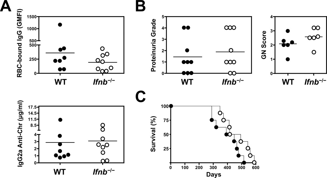
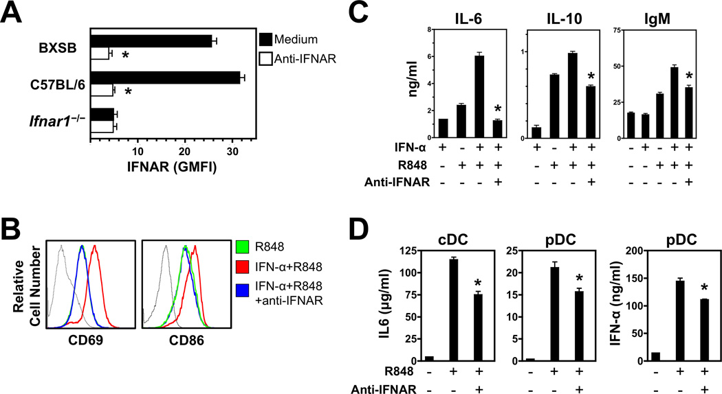
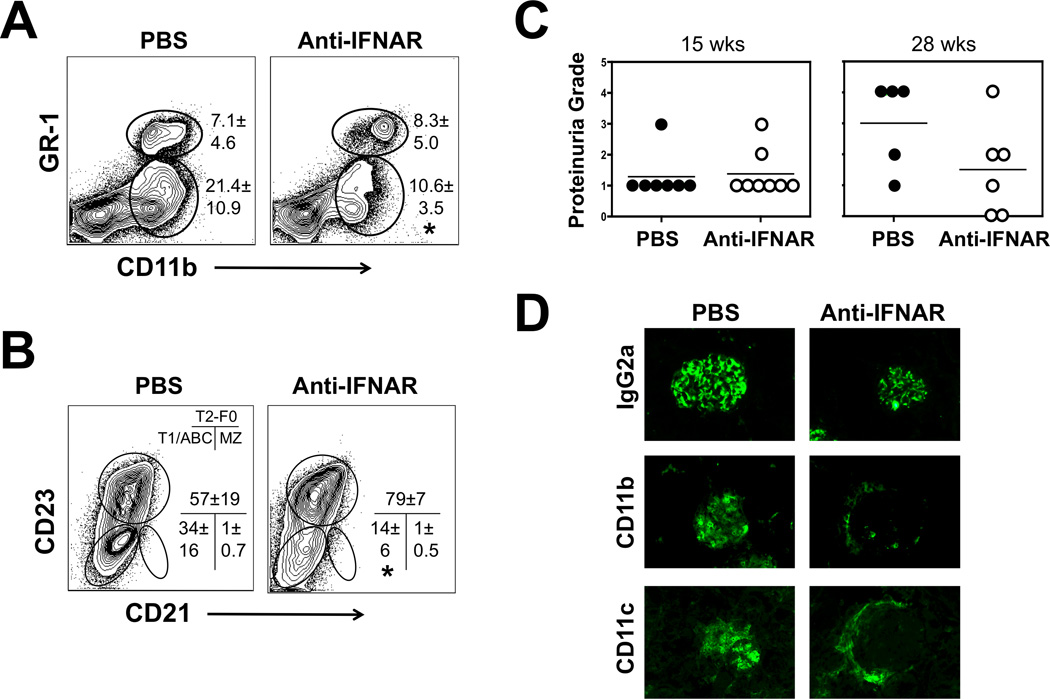
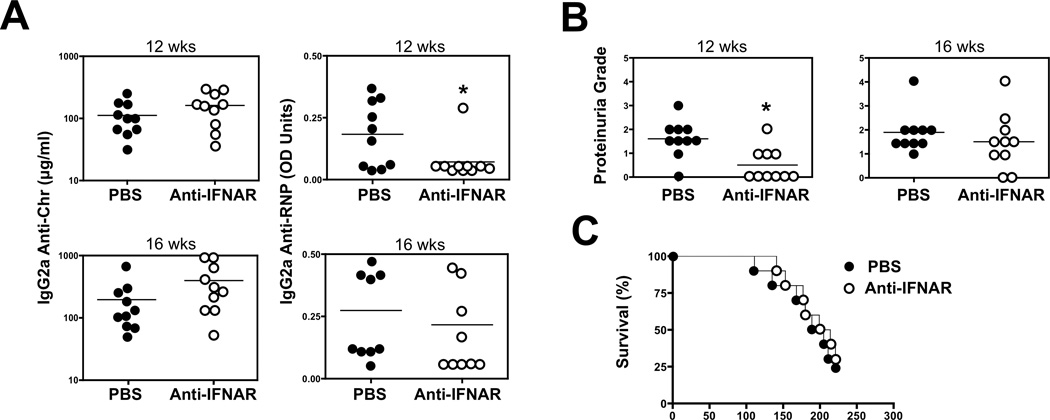
The demonstration in humans and mice that nucleic acid-sensing TLRs and type I IFNs are essential disease mediators is a milestone in delineating the mechanisms of lupus pathogenesis. In this study, we show that Ifnb gene deletion does not modify disease progression in NZB mice, thereby strongly implicating IFN-α subtypes as the principal pathogenic effectors. We further document that long-term treatment of male BXSB mice with an anti-IFN-α/β receptor Ab of mouse origin reduced serologic, cellular, and histologic disease manifestations and extended survival, suggesting that disease acceleration by the Tlr7 gene duplication in this model is mediated by type I IFN signaling. The efficacy of this treatment in BXSB mice was clearly evident when applied early in the disease process, but only partial reductions in some disease characteristics were observed when treatment was initiated at later stages. A transient therapeutic effect was also noted in the MRL-Fas(lpr) model, although overall mortality was unaffected. The combined findings suggest that IFN-α/β receptor blockade, particularly when started at early disease stages, may be a useful treatment approach for human systemic lupus erythematosus and other autoimmune syndromes.


Figures

Figure 1
Figure 1. IFN-β Is Not Required for Lupus-Like Disease in NZB Mice
Groups of Ifnb−/− and WT NZB mice (n = 8–9/group) were followed for disease manifestations. (A) Anti-red blood cells (RBC) and anti-chromatin autoantibody levels at 9 mo of age. (B) Proteinuria and glomerulonephritis scores at 10 mo of age. (C) Survival. p>0.05 for A–C.
Figure 2
Figure 2. IFNAR Expression in BXSB Mice
(A) Ex vivo analysis. Spleen cells from BXSB mice (16 wks old) were examined by flow cytometry to assess cell surface IFNAR expression after gating on T cells (CD4+ and CD8+), B cells (B220+), monocytes (CD11b+CD11c), conventional DCs (CD11c+CD11b+ and CD11c+CD11b), and pDCs (PDCA1+CD11clow). Gray-filled histograms indicate background staining. (B) IFNAR modulation. Splenic B cells from BXSB mice were cultured in vitro for 24 hrs with medium alone (control), or medium containing IFN-α, the TLR7 ligand R848, or both. IFNAR expression on gated B220+ cells (black-lined histograms) was examined as in panel A. Background staining (gray-lined histograms) and IFNAR levels in non-cultured ex vivo B cells (gray-filled histograms) are also depicted. Numbers in histograms represent geometric mean fluorescence intensity (GMFI) ± SD. Asterisks indicate statistical significance (p<0.05).
Figure 3
Figure 3. Anti-IFNAR Antibody Inhibits In Vitro Activation of B cells, DCs, and pDCs from BXSB Mice
(A–C) Effects of IFNAR blockade in B cells. (A) B cells from BXSB mice were incubated for 120 hrs in medium supplemented or not with anti-IFNAR antibody. Efficiency of IFNAR blockade on BXSB B cells (compared to similarly cultured B cells from WT and Ifnr1−/− C57BL/6 mice) was assessed by flow cytometry and expressed as GMFI ± SD. (B–C) B cells from BXSB mice were stimulated for 120 hrs with medium alone (control) or medium containing IFN-α, the TLR7 ligand R848, or both, in the presence or absence of anti-IFNAR antibody. The effect of IFNAR blockade on B cell activation was evaluated by measuring expression of CD69 and CD86 by flow cytometry (B) and production of IL-6, IL-10 and IgM by ELISA (C). (D) Effects of IFNAR blockade in DC subsets. Bone marrow-derived cDC and pDCs from BXSB mice were stimulated for 24 hrs with the TLR7 ligand R848 in the presence or absence of anti-IFNAR antibody. Production of IL-6 and IFN-α was determined by ELISA. Bars represent average (± SD) of individual mice. Asterisks indicate statistical significance (p<0.05).
Figure 4
Figure 4. Early anti-IFNAR Antibody Treatment of BXSB Mice
Mice (n = 8–10/group) were treated with anti-IFNAR antibody (or PBS) starting at 12 wks of age. (A) Efficiency of IFNAR blockade as defined 4 wks post-treatment initiation by flow cytometry (average GMFI ± SD) in T cells (CD4+ and CD8+), B cells (B220+), monocytes (MC, CD11b+CD11c), DCs (DC1, CD11c+CD11; DC2, CD11c+CD11b+), and pDCs (PDCA1+, CD11clow). Ifnar1−/− mice were used as negative staining controls (shown for B cells). (B) Serum autoantibody titers (anti-chromatin and ANA) at 20 wks of age. (C) Kidney histology showing glomerular size (PAS), immune deposits (IgG2a and complement C3), and immunocyte infiltration (CD11b+ and CD11c+ cells) at 38 wks of age (n = 3–4/group). (D) Glomerulonephritis scores at 38 wks of age (n = 3–4/group). (E) Survival. Arrow indicates start of antibody treatment. At the termination of the experiment (38 wks of age), three PBS-treated and five anti-IFNAR-treated mice were still alive. Asterisks indicate statistical significance (p<0.05).
Figure 5
Figure 5. Anti-IFNAR Antibody Treatment Inhibits Expansion of B cells, Monocytes, and Dendritic Cells in BXSB Mice
Mice (n = 8–10/group) were treated with anti-IFNAR antibody (or PBS) from 12 wks to 38 wks of age. (A) Weights (± SD) of spleen and lymph nodes (LN, inguinal, axillary and cervical). (B) B cell subsets. Spleen cells were examined for CD21 and CD23 expression after gating on B220+IgM+ B cells to identify CD21CD23+ T2/follicular (T2-FO), CD21+CD23 marginal zone (MZ), and CD21CD23 T1 immature and age-associated B cells (ABCs). Numbers within plots correspond to average frequency of the indicated subsets. Bar graph indicates cell numbers ± SD. (C) Expansion of CD21CD23AA4.1low B cells in untreated BXSB mice. Spleen cells were obtained from male BXSB mice (n = 5, age = 21 to 26 wks) with varying degrees of splenomegaly. The numbers of CD21CD23AA4.1low (ABCs) and CD21CD23AA4.1+ (T1) B cells were determined by flow cytometry and plotted as a function of the frequency of CD21CD23 B cells for each individual mouse. Linear regression was calculated using Prism 4 software. (D) Spleen monocytes (CD11b+CD11c) and DC subsets (CD11c+CD11b and CD11c+CD11b+). Frequencies and cell numbers (± SD) were defined by flow cytometry. (E) pDCs and TLR7 expression in spleen B cell follicles. Spleen sections were stained with fluorescent anti-PDCA1 (pDC, green) and anti-TLR7 (red), or with anti-CD19 (B cells, green) antibodies. Asterisks indicate statistical significance (p<0.05).
Figure 6
Figure 6. Late anti-IFNAR Antibody Treatment of BXSB Mice
Mice (n = 7–8/group) were treated with anti-IFNAR antibody starting at 17 wks of age. (A) Blood monocyte subsets (CD11b+Gr-1 "resident", CD11b+Gr-1+ "inflammatory") at 26 wks of age. Numbers indicate average frequencies (± SD) of the gated cell subsets (* = p < 0.05). (B) Spleen B cell subsets at 33 wks of age. Numbers indicate average frequencies (± SD) of the gated cell subsets (* = p < 0.05). (C) Proteinuria at 15 and 28 wks of age. (D) Kidney glomerular IgG2a deposits and immunocyte infiltration (CD11b+ and CD11c+) at 33 wks. Shown are representative immunofluorescence images of kidneys from treated and control mice (n=4/group). Scoring of individual mice (0–4, based on fluorescence intensity) indicated significant treatment-associated reductions in IgG2a deposits (2.25 ± 1.26 vs. 3.75 ± 0.5, p < 0.05), CD11b staining (1.13 ± 0.63 vs. 3.13 ± 1.75, p < 0.05) and CD11c staining (1.00 ± 0.82 vs. 2.5 ± 1.29, p < 0.05).
Figure 7
Figure 7. Prophylactic anti-IFNAR Antibody Treatment of MRL-Faslpr Mice
Mice (n = 10/group) were treated with anti-IFNAR antibody starting at 7 wks of age. (A) Serum anti-chromatin and anti-ribonucleoprotein (RNP) autoantibody titers. (B) Proteinuria. (C) Survival. Asterisks indicate statistical significance (p<0.05).

References

    1. Theofilopoulos AN, Baccala R, Beutler B, Kono DH. Type I interferons (alpha/beta) in immunity and autoimmunity. Annu Rev Immunol. 2005;23:307–335. - PubMed
    1. Banchereau J, Pascual V. Type I interferon in systemic lupus erythematosus and other autoimmune diseases. Immunity. 2006;25:383–392. - PubMed
    1. Ronnblom L, Eloranta ML, Alm GV. The type I interferon system in systemic lupus erythematosus. Arthritis Rheum. 2006;54:408–420. - PubMed
    1. Marshak-Rothstein A, Rifkin IR. Immunologically active autoantigens: the role of toll-like receptors in the development of chronic inflammatory disease. Annu Rev Immunol. 2007;25:419–441. - PubMed
    1. Baccala R, Hoebe K, Kono DH, Beutler B, Theofilopoulos AN. TLR-dependent and TLR-independent pathways of type I interferon induction in systemic autoimmunity. Nat Med. 2007;13:543–551. - PubMed

Publication types

MeSH terms