. 2012 Nov 21;32(47):16725-35a.
doi: 10.1523/JNEUROSCI.2153-12.2012.

Cognitive enhancement with rosiglitazone links the hippocampal PPARγ and ERK MAPK signaling pathways

Affiliations

Cognitive enhancement with rosiglitazone links the hippocampal PPARγ and ERK MAPK signaling pathways

Larry A Denner et al. J Neurosci. .

Abstract

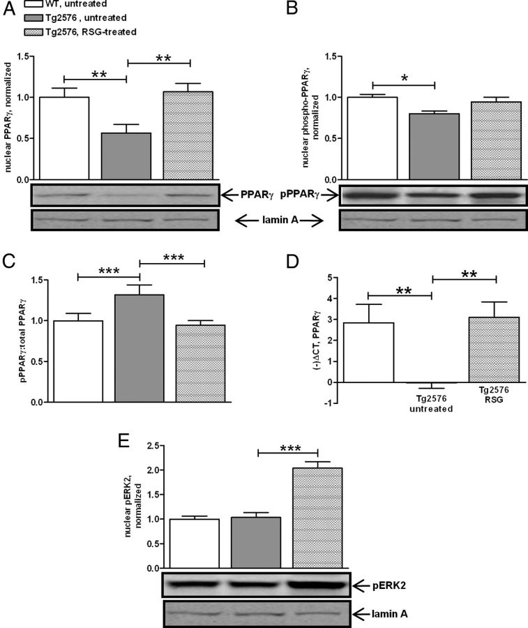
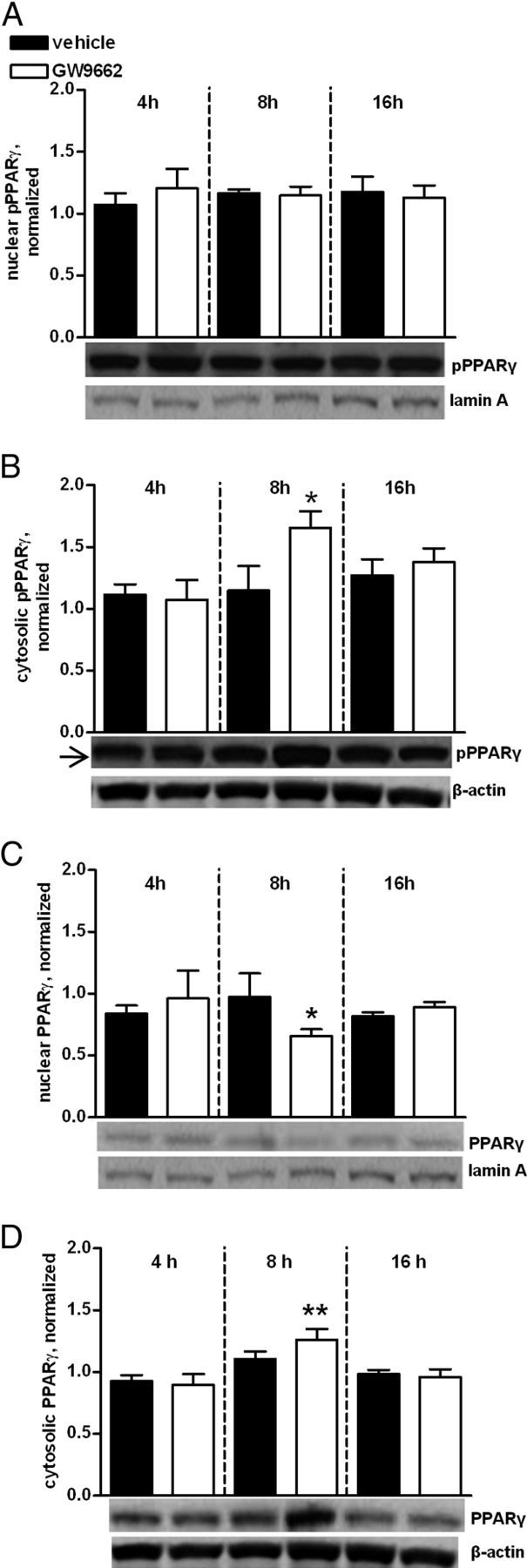
We previously reported that the peroxisome proliferator-activated receptor γ (PPARγ) agonist rosiglitazone (RSG) improved hippocampus-dependent cognition in the Alzheimer's disease (AD) mouse model, Tg2576. RSG had no effect on wild-type littermate cognitive performance. Since extracellular signal-regulated protein kinase mitogen-activated protein kinase (ERK MAPK) is required for many forms of learning and memory that are affected in AD, and since both PPARγ and ERK MAPK are key mediators of insulin signaling, the current study tested the hypothesis that RSG-mediated cognitive improvement induces a hippocampal PPARγ pattern of gene and protein expression that converges with the ERK MAPK signaling axis in Tg2576 AD mice. In the hippocampal PPARγ transcriptome, we found significant overlap between peroxisome proliferator response element-containing PPARγ target genes and ERK-regulated, cAMP response element-containing target genes. Within the Tg2576 dentate gyrus proteome, RSG induced proteins with structural, energy, biosynthesis and plasticity functions. Several of these proteins are known to be important for cognitive function and are also regulated by ERK MAPK. In addition, we found the RSG-mediated augmentation of PPARγ and ERK2 activity during Tg2576 cognitive enhancement was reversed when hippocampal PPARγ was pharmacologically antagonized, revealing a coordinate relationship between PPARγ transcriptional competency and phosphorylated ERK that is reciprocally affected in response to chronic activation, compared with acute inhibition, of PPARγ. We conclude that the hippocampal transcriptome and proteome induced by cognitive enhancement with RSG harnesses a dysregulated ERK MAPK signal transduction pathway to overcome AD-like cognitive deficits in Tg2576 mice. Thus, PPARγ represents a signaling system that is not crucial for normal cognition yet can intercede to restore neural networks compromised by AD.


Figures

Figure 1.
Figure 1.
Oral delivery of RSG impinges upon CNS PPARγ. A, Hippocampal PPARγ binding to its PPRE is enhanced by 1 month RSG treatment. Two-way ANOVA F(3,31) = 9.34 for treatment; no interaction was detected. B, One month RSG treatment induces PPARγ target gene expression. The mRNA for the PPRE-containing APO-O gene is reduced in untreated Tg2576 compared with WT untreated. RSG normalizes APO-O expression in Tg2576. One-way ANOVA of ΔCT values resulted in F(2,9) = 8.6. C, Quantitative mass spectrometry reveals Tg2576 hippocampal proteins altered with RSG treatment. All proteins displayed have a Benjamini-Hochberg rank sum p ≤ 0.05. D, Ingenuity Pathways Analysis of synaptic plasticity proteins identified by quantitative mass spectrometry placed ERK MAPK as a central node in the protein network. ANXA6, Annexin A6; CACNG8, voltage-dependent calcium channel γ-8 subunit; CPLX2, complexin 2; GAD1, glutamate decarboxylase 1; GR1A2, glutamate receptor subunit 2; GSK3A, glycogen synthase kinase-3α; MAP2K6, dual specificity mitogen-activated protein kinase kinase; PKA, protein kinase A; PPARG, PPARγ; PRKCG, protein kinase C-γ; RASAL1, RasGAP-activating-like protein 1; SIRPA, signal-regulatory protein α; SNCA, α-synuclein (see Materials and Methods and www.ingenuity.com for a more detailed description of network statistical calculations, molecule naming, and symbol descriptions). E, PCR strategy to detect PPARγ1 and PPARγ2 gene transcripts in mouse hippocampus. F, Both PPARγ1 and PPARγ2 are detected in hippocampus by conventional PCR (gel image, top). Quantitative PCR shows PPARγ1 mRNA expression is much higher than PPARγ2 in mouse hippocampus. *p < 0.05, **p < 0.01, ***p < 0.001.
Figure 2.
Figure 2.
RSG reverses deficits in nuclear PPARγ and increases nuclear ERK2 activity in hippocampus. A, Quantitative immunoblotting revealed significant downregulation of nuclear PPARγ in Tg2576 hippocampus. One-way ANOVA (F(2,23) = 7.02; p = 0.004). RSG treatment of Tg2576 normalized nuclear PPARγ to WT levels. B, Phosphorylation of nuclear PPARγ is decreased in Tg2576 and reversed with RSG treatment. One-way ANOVA, (F(2,16) = 3.2). C, The nuclear pPPARγ/total PPARγ ratio is increased in untreated Tg2576 compared with wild-type, and normalized with RSG. One-way ANOVA (F(2,16) = 19.4). D, RSG increases PPARγ gene expression. Quantitative PCR showed that PPARγ mRNA was reduced in untreated Tg2576 and normalized to WT levels with RSG treatment. One-way ANOVA resulted in (F(2,9) = 8.2). E, Hippocampal nuclear pERK2 levels are equivalent between untreated WT and untreated Tg2576 but increased in RSG-treated Tg2576. One-way ANOVA (F(2,17) = 37.3) and Dunnett's post hoc analysis. Data reported normalized to untreated WT; mean ± SEM. *p < 0.05, **p < 0.01, ***p < 0.001.
Figure 3.
Figure 3.
Inhibition of CNS PPARγ blocks RSG-mediated cognitive rescue. Untreated or RSG-treated mice were infused with either vehicle or GW9662 4 h before 2-pairing FC training. A, Timeline for FC training and testing following ICV infusion of GW9662. ICV injection was performed 4 h before the acquisition of FC learning (FC Training). Consolidation proceeds for up to ∼10 h following FC training. Testing for recall of FC 24 h after training tests for consolidation of FC learning. B, No genotype or treatment effects were detected in the 2-pairing training for FC. Repeated-measures two-way ANOVA (F(1,1,1) = 2.49 and 2.00) for genotype and treatment, respectively; no interaction was detected. Data reported as mean percentage freezing ± SEM for each 30 s epoch. Vertical arrows on timeline denote the epoch within which the footshock was delivered during FC training. C, In the contextual test for FC, two-way ANOVA detected a genotype effect but no treatment effect or interaction (F(2,1,2) = 0.778 and 29.72) for genotype and treatment. Therefore, untreated Tg2576 (RSG−) vehicle-infused (GW V) Tg2576 and RSG-treated (RSG+) Tg2576 ICV infused with GW9662 (GW+) froze significantly less. Neither RSG nor GW9662 had an effect on performance of WT. Data reported as mean percentage total freezing ± SEM. ***p < 0.0001 compared with RSG-vehicle groups; **p < 0.01 compared with vehicle-infused groups. V, Vehicle-infused. D, No significant genotype or treatment effect detected in 9MO WT and Tg2576, untreated or RSG-treated, with two-way ANOVA in the shock threshold test.
Figure 4.
Figure 4.
Hippocampal nuclear ERK2 activity is modulated by PPARγ. A–D, Quantitative immunoblot of hippocampal total and pPPARγ in nuclear and cytoplasmic compartments from RSG-treated Tg2576 ICV infused with vehicle or GW9662. ICV injection of GW9662 analyzed by one-way ANOVA detected no effect on nuclear pPPARγ at any time point (A) (F(6,20) = 0.49), but did result in a significant increase in cytosolic pPPARγ by 8 h (B) (F(6,24) = 3.16). C, D, One-way ANOVA and Dunnett's post hoc analysis revealed that ICV injection of GW9662 led to a significant decrease in nuclear PPARγ levels 8 h after infusion (C), with a concomitant increase in cytosolic PPARγ (D) (F(6,30) = 2.83 and 3.38) for C and D, respectively. Data normalized to RSG-treated Tg2576 and expressed as mean ± SEM. *p < 0.05, **p < 0.01, ***p < 0.001.

References

    1. Ahi J, Radulovic J, Spiess J. The role of hippocampal signaling cascades in consolidation of fear memory. Behav Brain Res. 2004;149:17–31. - PubMed
    1. Alessi DR, Gomez N, Moorhead G, Lewis T, Keyse SM, Cohen P. Inactivation of p42 MAP kinase by protein phosphatase 2A and a protein tyrosine phosphatase, but not CL100, in various cell lines. Curr Biol. 1995;5:283–295. - PubMed
    1. Altiok S, Xu M, Spiegelman BM. PPARgamma induces cell cycle withdrawal: inhibition of E2F/DP DNA-binding activity via down-regulation of PP2A. Genes Dev. 1997;11:1987–1998. - PMC - PubMed
    1. Atkins CM, Selcher JC, Petraitis JJ, Trzaskos JM, Sweatt JD. The MAPK cascade is required for mammalian associative learning. Nat Neurosci. 1998;1:602–609. - PubMed
    1. Bell KA, O'Riordan KJ, Sweatt JD, Dineley KT. MAPK recruitment by beta-amyloid in organotypic hippocampal slice cultures depends on physical state and exposure time. J Neurochem. 2004;91:349–361. - PubMed

Publication types

MeSH terms

LinkOut - more resources