. 2012 Nov 21;32(47):16857-71a.
doi: 10.1523/JNEUROSCI.1858-12.2012.

The complex PrP(c)-Fyn couples human oligomeric Aβ with pathological tau changes in Alzheimer's disease

Affiliations

The complex PrP(c)-Fyn couples human oligomeric Aβ with pathological tau changes in Alzheimer's disease

Megan Larson et al. J Neurosci. .

Retraction in

Abstract

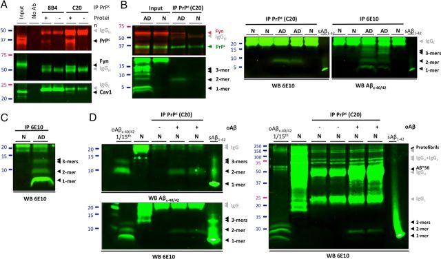
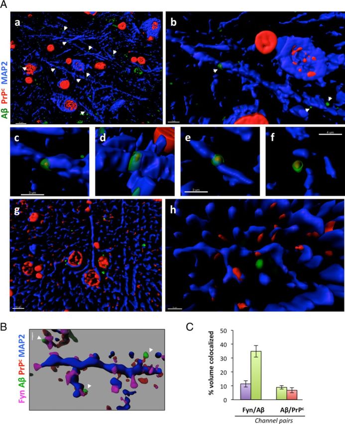
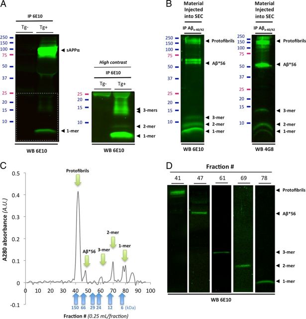
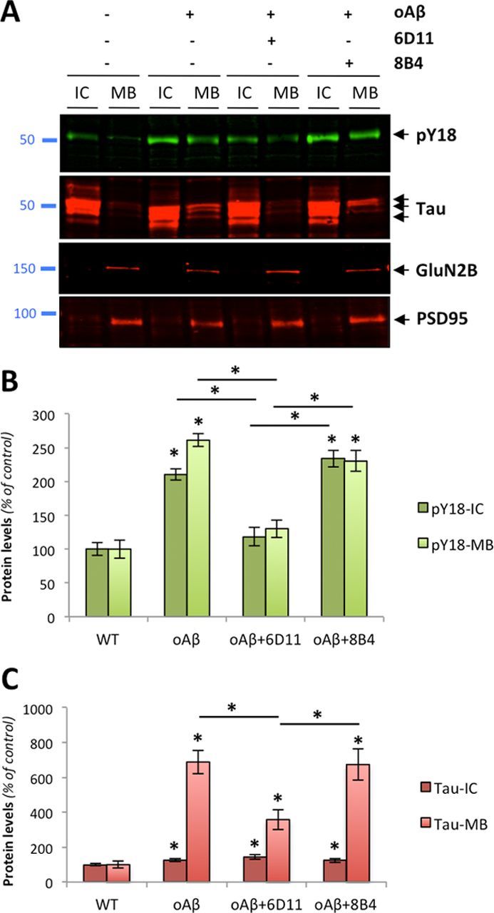
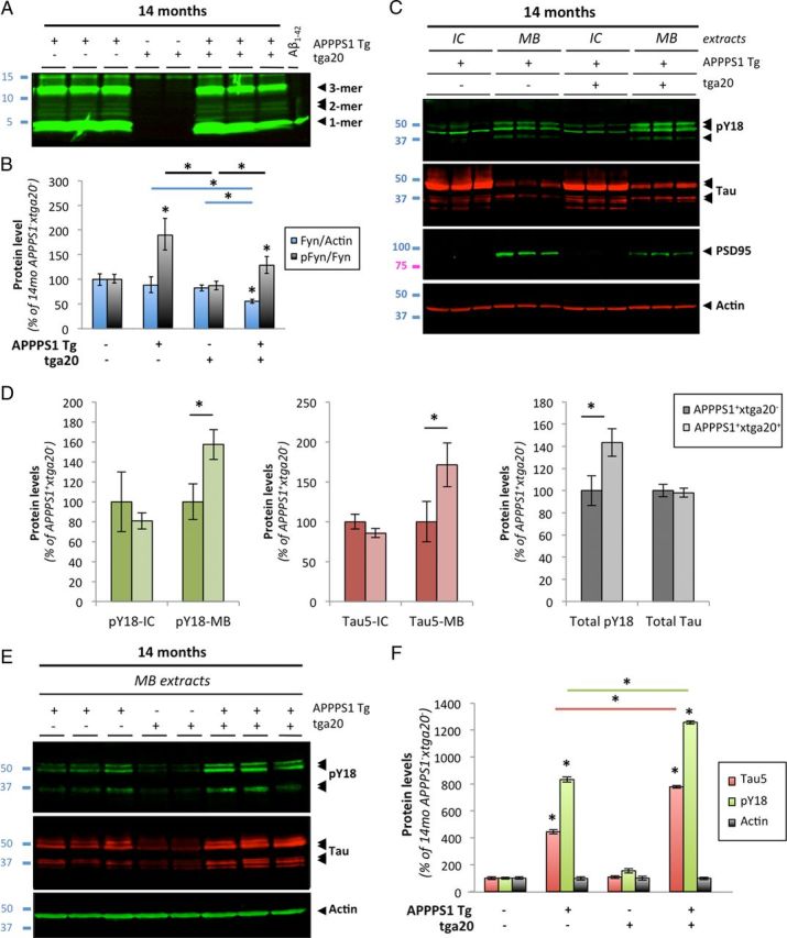
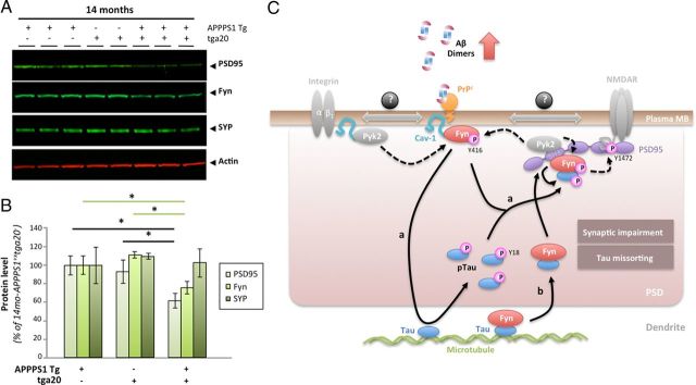
Amid controversy, the cellular form of the prion protein PrP(c) has been proposed to mediate oligomeric amyloid-β (Aβ)-induced deficits. In contrast, there is consistent evidence that the Src kinase Fyn is activated by Aβ oligomers and leads to synaptic and cognitive impairment in transgenic animals. However, the molecular mechanism by which soluble Aβ activates Fyn remains unknown. Combining the use of human and transgenic mouse brain tissue as well as primary cortical neurons, we demonstrate that soluble Aβ binds to PrP(c) at neuronal dendritic spines in vivo and in vitro where it forms a complex with Fyn, resulting in the activation of the kinase. Using the antibody 6D11 to prevent oligomeric Aβ from binding to PrP(c), we abolished Fyn activation and Fyn-dependent tau hyperphosphorylation induced by endogenous oligomeric Aβ in vitro. Finally, we showed that gene dosage of Prnp regulates Aβ-induced Fyn/tau alterations. Together, our findings identify a complete signaling cascade linking one specific endogenous Aβ oligomer, Fyn alteration, and tau hyperphosphorylation in cellular and animal models modeling aspects of the molecular pathogenesis of Alzheimer's disease.


Figures

Figure 1.
Figure 1.
Increased membrane-bound PrPc levels are associated with Fyn activation in AD. A, Representative Western blot (WB) for PrPc, Fyn, pY416-Fyn, and NeuN in 10 of 84 specimens of NCI (N), MCI (M), and AD (A) composing our cohort. PrPc, Fyn, and α-tubulin were measured by direct WB, while pY416-Fyn levels were estimated following total Fyn immunoprecipitation. B–D, Box plots for PrPc (B), Fyn (C), and pY416-Fyn (D) protein levels in the MB fraction of NCI, MCI, and AD groups. Total Fyn levels were unchanged, whereas the levels of its active phosphorylated form, pY416-Fyn, were increased in the AD group compared with NCI and MCI [Kruskal–Wallis followed by Mann–Whitney's U test and Bonferroni's correction (p < 0.05)]. E, F, Linear regression analyses between PrPc/Fyn (E) and PrPc/pY416-Fyn (F) protein levels (Spearman's rank correlation). PrPc and total Fyn showed a strong association regardless of clinical status (E). In contrast, a significant positive correlation was only found in the AD group linking activated Fyn and PrPc levels. In the figure legends, the italicized numbers between parentheses indicate group sizes. NCI is shown in green, MCI in yellow/orange, and AD in red. In box plots, the bar inside the box indicates the median; the upper and lower limits of boxes represent the 75th and 25th percentiles, respectively; and the bars flanking the box represent 95th and 5th percentiles. *p < 0.05; **p < 0.01. EC, extracellular-enriched fraction; IC, intracellular-enriched fraction; MB, membrane-associated fraction; Tg, transgenic; DLU, densitometry light unit; A.U., arbitrary unit.
Figure 2.
Figure 2.
PrPc immunoprecipitates specifically with human soluble Aβ dimers. A, Potential interaction of PrPc with Fyn in AD brain tissues was assessed by SDS-PAGE analysis of the PrPc/caveolin-1/Fyn complex following immunoprecipitation of PrPc (with 8B4 or C20 antibodies) or Fyn. The gray arrowheads indicate exogenous antibodies used for IP. B, PrPc forms a putative complex with Fyn and soluble Aβ dimers using AD brain tissue. Western blots for Aβ were performed with 6E10. Following membrane stripping, PrPc and Fyn were revealed with PrP C20 and Fyn3 antibodies. Additional IPs with different cases are shown on the right panels to illustrate the reproducibility of the findings. C, Western blot analysis of TBS-soluble extracts from selected brains of subjects with AD or no cognitive impairment (N). Monomers, dimers, and trimers are readily detected using 6E10. D, Immunoprecipitation of PrPc with exogenous human Aβ dimers in a cell-free assay. Affinity-purified human soluble Aβx-40/42 species (lane 1, left and right panels, an estimated total of 2.85 ng relative to Aβ1–42 standards) were added to brain protein extracts with no detectable Aβ (N; same as A); PrPc was immunoprecipitated using the C20 antibody and bound Aβ species were detected with 40/42-end specific antibodies (top left panel) or 6E10 (bottom left and right panels). Full Western blot images indicated that isolated large soluble Aβ assemblies (e.g., Aβ*56 and protofibrils) were not pulled down by C20 in our assay (right panel). IP, immunoprecipiration; No Ab, no antibody; N, nonimpaired age-matched control brain; AD, Alzheimer's disease brain; oAβ, endogenous oligomeric Aβ species purified from human AD brain tissue; sAβ1–42, 0.5 ng of human synthetic Aβ1–42 (Sigma-Aldrich).
Figure 3.
Figure 3.
PrPc and soluble Aβ colocalize at dendritic spines in human AD brain tissues. Surface rendering of triple-channel confocal immunofluorescence reveals that PrPc (in red, labeled using the C20 antibody) and Aβ (green, labeled using the DW6 antiserum) colocalize with the postsynaptic protein Fyn (magenta) along neuronal dendritic shafts (in blue, labeled using a MAP-2 antibody) in human brain tissues [AD (a–f); age-matched NCI (g, h)]. Images were acquired using oil-immersion 60 or 100× objectives and processed with Imaris7.0 software. Transparency of the 405, 488, 536 nm channels (magenta, green, red) was increased to 40% to visualize the rendered volumes of all other fluorescent probes conjointly. A, Low- and high-power images of PrPc/Aβ puncta along dendritic shafts of inferior temporal gyrus neurons. Examples of colocalization between PrPc and Aβ at higher magnification are shown in b–f. Please note the lack of colocalization of PrPc and Aβ on dendrites of NCI brain sections (g, h). Scale bars: a, g, 10 μm; b–e, 3 μm; f, 4 μm; h, 5 μm. B, PrPc:Aβ complexes colocalize with the dendritic spine protein Fyn in vivo. Scale bar, 1 μm. C, Software-assisted quantification of colocalized voxels (N = 3 subjects/6 sections/18 ROIs). Bars represent the mean ± SD.
Figure 4.
Figure 4.
Biochemical characterization of soluble Aβ species present in Tg2576 conditioned medium and in human brain tissues. Western blot analyses of affinity-purified soluble Aβ species present in conditioned media (CM) of Tg2576 primary cortical neurons and in soluble TBS extracts human AD brain. A, Immunoprecipitation of soluble APP/Aβ molecules present in CM of Tg2576 primary neurons (DIV12–14) using 6E10. A high-intensity laser scan is also shown to better visualize Aβ monomers, dimers, and trimers (bottom inset). B, Representative profile of soluble Aβ oligomers affinity-purified with 40- and 42-end specific antibodies (Mab 2.1.3 and Mab13.1.1; kindly provided by Dr. Pritam Das) to prevent capturing APP molecules and detected with 6E10 (left panel) or 4G8 (right panel). Several soluble Aβ assemblies are readily observed: monomers, dimers, trimers, Aβ*56, a larger 60 kDa oligomer, and protofibrils >150 kDa. C, Typical size exclusion chromatogram of the endogenous human Aβ oligomers shown in B using a Tricorn Superdex 75 column. Five peaks (green arrows) corresponding to the five Aβ species seen by Western blot are clearly observed with elutions at predicted molecular weights (blue arrows correspond to the six molecular weight standards used). D, Representative Western blot images of the respective size exclusion chromatography fractions isolated in C using 6E10.
Figure 5.
Figure 5.
Specific PrPc antibodies prevent soluble Aβ-induced Fyn activation in primary neurons. A, Quadruple-channel confocal immunofluorescence reveals that PrPc (labeled using the C20 antibody) and Aβ (labeled using the DW6 antiserum, a kind gift from Dr. Dominic Walsh) colocalize on neuronal dendritic spines (labeled using a PSD95 antibody) in 14-d-old primary cortical Tg2576 neurons. B, Quantification of colocalized voxels for each channel pair of interest. Representative 3D-rendered images used for voxel counts are shown in the left panel. Results for channel pairs are shown in the histogram. C, Triple-channel confocal imaging shows that Aβ42 species partly colocalize with pY416-Src and PrPc in Tg2576 primary neurons. D, Exogenous Aβ species purified from AD brain tissue colocalized with PrPc and Fyn at dendritic spines after application onto nontransgenic primary cortical neurons. E, F, Fyn activation induced by human oligomeric Aβ is partially blocked by the antibody 6D11 targeting the 95–105 domain of PrPc. Preincubation with the N terminus PrPc antibody 8B4 failed to inhibit Fyn phosphorylation. 6D11 (10 μg) and 8B4 (10 μg) were preincubated for 2 h onto cells before acute oAβ exposure (1 h at 37°C). 6D11 reduced Fyn activation in wild-type neurons acutely exposed to human-brain purified Aβ oligomers (E) and in Tg2576 neurons chronically exposed to low-n Aβ oligomers (F). Images were acquired using an oil-immersion 60 or 100× objective and processed with Imaris7.1 software. The arrowheads indicate examples of colocalization between Aβ/PrPc, while white asterisks correspond to colocalization between Aβ and PSD95 in absence of PrPc. Bars represent the mean ± SD (n > 3–6 dishes/experiment; independent experiments were performed in triplicate). *p < 0.05, ANOVA followed by Student's t test.
Figure 6.
Figure 6.
PrPc-dependent phosphorylation of tau induced by endogenous Aβ oligomers in primary neurons. A, Protein levels of hyperphosphorylated tau at Y18 (pY18-Tau) and total tau as assessed by Western blot using PY18 and Tau5 antibodies in wild-type primary neurons exposed to human-brain purified oligomeric Aβ (5 nm for 1 h). The glutamatergic NMDA receptor subunit 2B (GluN2B) and the scaffolding protein PSD95 were used as internal control. B, C, Quantitation of pY18-Tau (B) and total tau (C) across conditions and protein fractions (IC and MB) revealed a significant increase in tau hyperphosphorylation and mislocalization following oAβ exposure. This effect was partially inhibited by 6D11 but not 8B4 (bars represent the mean ± SD; *p < 0.05, ANOVA followed by Student's t test).
Figure 7.
Figure 7.
Human AD brain-purified Aβ dimers and trimers activate Fyn in primary cortical neurons. A, Application of purified soluble Aβ dimers and trimers selectively phosphorylate Fyn and tau as assessed by quantitative Western blotting (duplicates are shown; n = 4 per experiment). Sixty-minute-long treatments were performed at the calculated concentration of 5 nm for all species (estimated based on monomeric synthetic Aβ levels). Total protein levels of GluN2B and phosphorylation of GluN2B at Y1472 were also examined. Actin levels are shown as control. B, Quantification of the ratios for pY416-Src/Fyn (labeled as pFyn/Fyn), pY18-Tau/total tau (pTau/Tau), and pY1472-GluN2B/GluN2B (pGluN2B/GluN2B) after 1-h-long exposure with the human oligomeric Aβ species listed. Both brain-purified Aβ dimers and trimers activate Fyn in vitro. Isolated monomeric Aβ, Aβ*56, and soluble Aβ PFs did not induce Fyn phosphorylation under similar conditions. After 60 min, neither membrane-bound levels of GluN2B nor Y1472-GluN2B phosphorylation were altered by AD brain-derived Aβ species (bars represent the mean ± SD; *p < 0.05, ANOVA followed by Student's t test with Bonferroni's correction).
Figure 8.
Figure 8.
Prnp gene ablation attenuates Aβ-induced activation of Fyn and tau hyperphosphorylation in vivo. Gene ablation of Prnp modulates Fyn activation and tau phosphorylation at pY18 in brains of aged transgenic APPPS1+×Prnp animals. A, Protein levels of low-molecular-weight Aβ oligomers, total Fyn, and pY416-Src were assessed by quantitative Western blot (WB). 6E10 was used for Aβ. A marked reduction of pY416 was revealed in APPPS1+×Prnp−/− compared with APPPS1+×Prnp+/− mice. B, Relative protein levels for total Fyn and pY416-Fyn determined by densitometry analyses. C, D, Prnp gene deletion limits tau hyperphorphorylation and missorting in vivo. PY18-tau and total tau levels were measured by WB using either intracellular-enriched or membrane-enriched protein extracts (C) and quantified by software analysis (D). APPPS1+×Prnp−/− mice showed a more than twofold reduction in tau phosphorylation at Y18 compared with APPPS1+×Prnp+/− littermates (bars represent the mean ± SD; *p < 0.05, ANOVA followed by Student's t test; n ≥ 3 animals/genotype/age/experiment).
Figure 9.
Figure 9.
Aβ-induced activation of Fyn and tau hyperphosphorylation is enhanced in aged APPS1 mice overexpressing PrP. A, Soluble brain levels of low-molecular-weight Aβ oligomers in 14-month-old APPPS1×tga20 mice was assessed by quantitative Western blot (WB) using 6E10. B, Quantification of total Fyn, pY416-Src, and actin protein levels in 14-month-old mice (APPPS1×tga20, APPPS1+×tga20, APPPS1×tga20+, and APPPS1+×tga20+) following WB. A marked elevation of pY416-Fyn was observed in APPPS1+×tga20+ compared with APPPS1×tga20+ mice. C, Tau missorting was illustrated by biochemical segregation of pY18-Tau, total tau, PSD95 in IC versus MB extracts using Western blotting. D, Quantification of PY18-tau and total tau immunoreactivity across protein fractions and genotypes. Aged APPPS1+×tga20+ mice showed a ∼1.8-fold accumulation of tau and a ∼1.6-fold increase in phosphorylation at Y18 compared with age-matched APPPS1+×tga20 mice. E, Enhanced accumulation of full-length tau and cleavage products phosphorylated at Y18 in PSD95-containing extracts of old APPPS1+×tga20+ mice. Actin was used as internal standard. F, Quantification of tau species immunoreactive for Tau5 and PY18 in 14-month-old APPPS1×tga20, APPPS1+×tga20, APPPS1×tga20+, and APPPS1+×tga20+ mice (bars represent the mean ± SD; *p < 0.05, ANOVA followed by Student's t test; n ≥ 3 animals/genotype/age/experiment).
Figure 10.
Figure 10.
PrPc overexpression in aged APPPS1 mice triggers a selective decrease in postsynaptic proteins. A, Apparent selective decrease in postsynaptic proteins PSD95 and Fyn in 14-month-old APPPS1+×tga20+ mice. Representative WBs for the postsynaptic proteins PSD95 and Fyn as well as for the presynaptic vesicle protein synaptophysin (SYP) are shown. Actin was used as internal standard. B, Quantification of PSD95, Fyn, and Synaptophysin in 14-month-old APPPS1+×tga20, APPPS1×tga20+, and APPPS1+×tga20+ mice revealed significant reductions in postsynaptic proteins in APPPS1 mice overexpressing PrPc (bars represent the mean ± SD; *p < 0.05, ANOVA followed by Student's t test; n ≥ 3 animals/genotype/age/experiment). C, Proposed model of tau regulation by the triad oAβ-PrPc-Fyn. In the presence of accumulating Aβ dimers, PrPc, Fyn, and Cav-1 form a complex at the plasma membrane (likely with receptors such GluN or integrins known to interact with Cav-1 and Fyn). Upon phosphorylation of Fyn at Y416 (possibly by the kinase Pyk2), this complex becomes biologically active. Two scenarios are possible depending on the status of Fyn with respect to tau: Fyn is not bound to tau (a), and Fyn is bound to tau (b). In a, activated Fyn causes the hyperphosphorylation of tau at Y18 and its aberrant accumulation at the postsynaptic density (PSD). It is possible that pFyn might be bound to pTau at this stage and binds the SH3 domain of PSD95, where it is ideally located to modulate GluN/NMDA receptor subunits (i.e., GluN2B at Y1472). In model b, Fyn is already bound to tau in the dendrite, translocate to the PSD to interact with PSD95. There, Pyk2 could phosphorylate Fyn at Y416, resulting in Fyn activation and tau phosphorylation at Y18.

References

    1. State of aggregation. Nat Neurosci. 2011;14:399. [No authors listed] - PubMed
    1. Balducci C, Beeg M, Stravalaci M, Bastone A, Sclip A, Biasini E, Tapella L, Colombo L, Manzoni C, Borsello T, Chiesa R, Gobbi M, Salmona M, Forloni G. Synthetic amyloid-β oligomers impair long-term memory independently of cellular prion protein. Proc Natl Acad Sci U S A. 2010;107:2295–2300. - PMC - PubMed
    1. Barry AE, Klyubin I, Mc Donald JM, Mably AJ, Farrell MA, Scott M, Walsh DM, Rowan MJ. Alzheimer's disease brain-derived amyloid-β-mediated inhibition of LTP in vivo is prevented by immunotargeting cellular prion protein. J Neurosci. 2011;31:7259–7263. - PMC - PubMed
    1. Benilova I, De Strooper B. Prion protein in Alzheimer's pathogenesis: a hot and controversial issue. EMBO Mol Med. 2010;2:289–290. - PMC - PubMed
    1. Bennett DA, Wilson RS, Schneider JA, Evans DA, Beckett LA, Aggarwal NT, Barnes LL, Fox JH, Bach J. Natural history of mild cognitive impairment in older persons. Neurology. 2002;59:198–205. - PubMed

Publication types

MeSH terms