. 2012 Nov 21;32(47):16959-70.
doi: 10.1523/JNEUROSCI.1888-12.2012.

KAYAK-α modulates circadian transcriptional feedback loops in Drosophila pacemaker neurons

Affiliations

KAYAK-α modulates circadian transcriptional feedback loops in Drosophila pacemaker neurons

Jinli Ling et al. J Neurosci. .

Abstract

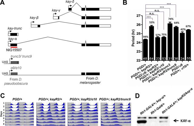
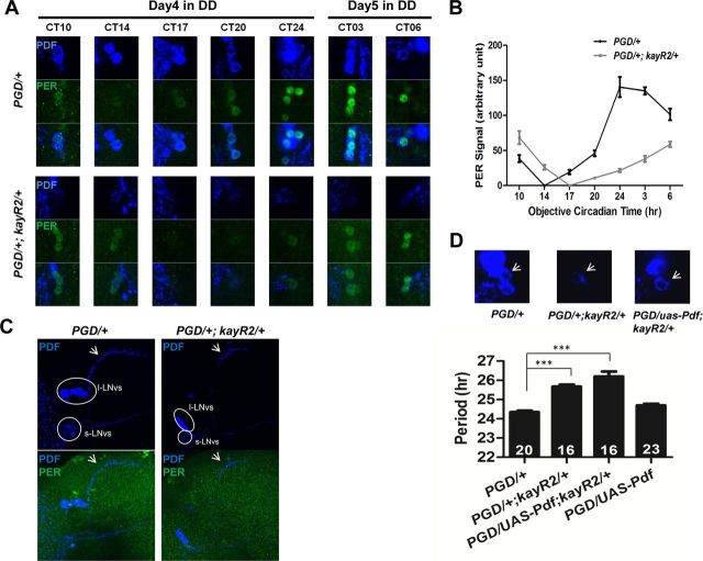
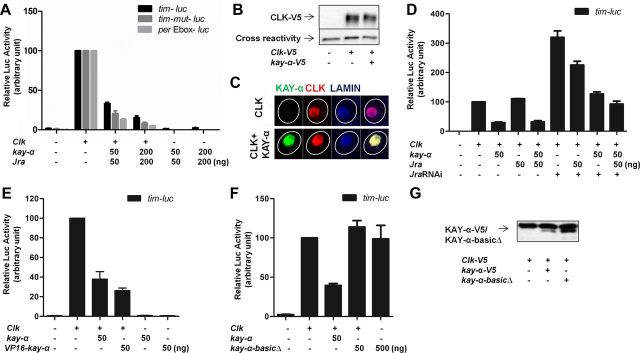
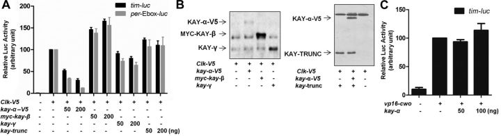
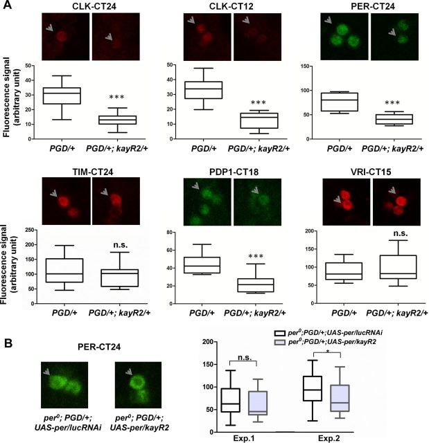
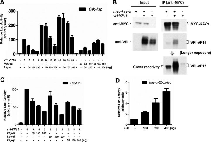
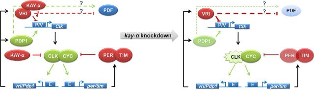
Circadian rhythms are generated by well-conserved interlocked transcriptional feedback loops in animals. In Drosophila, the dimeric transcription factor CLOCK/CYCLE (CLK/CYC) promotes period (per), timeless (tim), vrille (vri), and PAR-domain protein 1 (Pdp1) transcription. PER and TIM negatively feed back on CLK/CYC transcriptional activity, whereas VRI and PDP1 negatively and positively regulate Clk transcription, respectively. Here, we show that the α isoform of the Drosophila FOS homolog KAYAK (KAY) is required for normal circadian behavior. KAY-α downregulation in circadian pacemaker neurons increases period length by 1.5 h. This behavioral phenotype is correlated with decreased expression of several circadian proteins. The strongest effects are on CLK and the neuropeptide PIGMENT DISPERSING FACTOR, which are both under VRI and PDP1 control. Consistently, KAY-α can bind to VRI and inhibit its interaction with the Clk promoter. Interestingly, KAY-α can also repress CLK activity. Hence, in flies with low KAY-α levels, CLK derepression would partially compensate for increased VRI repression, thus attenuating the consequences of KAY-α downregulation on CLK targets. We propose that the double role of KAY-α in the two transcriptional loops controlling Drosophila circadian behavior brings precision and stability to their oscillations.


Figures

Figure 1.
Figure 1.
Downregulating kay-α lengthens the period of free-running circadian behavior in DD. A, Organization of the kay locus in D. melanogaster. kay is predicted to produce five isoforms. The dark boxes indicate coding sequences, and open boxes indicate noncoding sequences. Transgene NIG15507 generates dsRNAs targeting both the α and trunc isoforms. The red line indicates the region targeted by dsRNA. Constructs for cross-species rescue experiments are shown on the bottom of the panel. α9, α10, trunc3, and trunc9 are insertions of UAS-controlled transgenes that generate kay mRNAs resistant to the NIG15507 dsRNAs. The region targeted by NIG15507 dsRNAs was replaced with homologous D. pseudoobscura sequences. B, Downregulation of kay-α lengthens circadian behavior period. Bars 1 and 2, Flies expressing dsRNAs targeting kay-α and kay–trunc under the Pdf–GAL4 driver have ∼26-h-long period rhythms (control, 24.4 h). PGD: Pdf-GAL4, UAS-dcr2. Bars 3–6, The kay RNAi phenotype can be rescued with the kay-α construct resistant to the dsRNAs but not with the kay–trunc construct. Bars 7 and 8, The rescue is not explained by a period shortening caused by expression of the chimeric kay-α, because its expression in wild-type flies does not shorten circadian behavioral rhythms (it actually slightly lengthens them). Error bars correspond to SEM. Digits in the bar are the numbers of tested flies. Percentage of rhythmicity is indicated above the bars. One-way ANOVA, p < 0.0001. Tukey's multiple comparison test. ***p < 0.001; n.s., not significant at level of 0.05. C, Double-plotted actograms showing the average activity for each genotype. Flies were entrained in standard LD cycle for 3 d and then released in DD. D, The NIG15507-R2 transgene can inhibit KAY-α expression in vivo. Fly head extracts were immunoblotted with an anti-KAY-α/TRUNC antibody. Strong immunoreactivity was observed when KAY-α was misexpressed in the eyes with the Rh1–GAL4 driver (lane 1), whereas endogenous KAY-α was undetectable (lane 2). dsRNAs generated by NIG15507-R2 transgene were able to dramatically knock down the overexpression of KAY-α (lane 3).
Figure 2.
Figure 2.
Altered PER rhythms and reduced PDF levels in pacemaker neurons of kay-αRNAi flies. A, Confocal images of brains from control and kay-αRNAi (PGD/+; kayR2/+) flies immunostained with PER and PDF antibodies. Flies were entrained to a LD cycle for 3 d and then released in DD. Fly brains were dissected at indicated CTs during the fourth subjective night and fifth subjective day. Representative sLNvs are shown. B, Quantification of PER signals after subtraction of background signal. At CT14 for control flies and CT17 for kay-αRNAi flies, PER signals are indistinguishable from background; thus, they are set to “0” on the plot. Error bars correspond to SEM. C, The sLNvs develop normally in kay-αRNAi flies. Fly brains were dissected at CT24 on the first day in DD after 3 d of standard LD cycle and were immunostained for PDF and PER. Images are Z-stack projections of confocal images. Neuronal processes from the sLNvs to the dorsal brain (arrows) appear indistinguishable between PGD/+ and PGD/+; kayR2/+ flies in morphology. Circled are the regions containing cell bodies of large or small LNvs. PGD/+; kayR2/+ flies have only very weak PDF staining in the cell bodies of sLNvs. D, Restoring PDF levels does not rescue the long period phenotype. Overexpressing Pdf in kay-αRNAi flies restored PDF levels in sLNvs (the third panel, white arrow) but did not rescue the long period phenotype (bar 3), which is thus not attributable to low PDF levels but to a defective pacemaker. One-way ANOVA, p < 0.0001. Tukey's multiple comparison test. ***p < 0.001.
Figure 3.
Figure 3.
KAY-α represses CLK/CYC transactivation. Drosophila S2R+ cells were transfected as indicated. Luciferase activity was measured 2 d after transfection. A β-galactosidase-expressing plasmid was cotransfected to normalize transfection efficiency. The relative luciferase activity with Clk was set to 100 on each graph. A, KAY-α represses CLK activation of the tim promoter and the per E-box. The proximal tim promoter with a wild-type or mutagenized AP-1 binding site and multimerized per E-boxes were cloned in the pGL3 luciferase reporter vector to make tim–luc, tim–mut–luc, and per–Ebox–luc. Jra was cotransfected with kay-α. B, KAY-α does not alter CLK expression level in S2R+ cells. Cell lysates were immunoblotted with anti-V5 antibody. CLK–V5 protein level was quantified. CLK–V5 levels did not change whether or not kay-α was transfected in three independent experiments. CLK-V5 levels in the absence of KAY-α were set at 100. In the presence of KAY-α, relative CLK amount was 108 ± 20, n = 3. C, KAY-α does not alter CLK subcellular localization in S2R+ cells. Representative immunostaining showing CLK localization in the nucleus in the presence or absence of KAY-α. More than 95% of cells expressing or not KAY-α show this primarily nuclear CLK localization. Very rarely, and in both cells with or without KAY-α, CLK showed both nuclear and cytoplasmic localization. Lamin stains inner nuclear membrane. Circles outline the cell bodies. D, The repression of CLK activation by KAY-α does not require JRA. S2R+ cells were treated with dsRNAs targeting Jra 1 d before transfection to knock down endogenous JRA. Even in the presence of these Jra dsRNAs, KAY can still repress CLK/CYC activation. E, VP16–KAY-α cannot activate the proximal tim promoter. The activation domain of VP16 was fused to the N terminal of KAY-α to generate VP16–KAY-α. The fusion protein could not activate tim–luc but still repressed the activity of CLK. F, KAY-α DNA binding domain is required for CLK repression. The basic region of KAY-α was deleted to generate a kay-α–basicΔ construct, which was not able to repress CLK activity. G, The removal of the DNA binding domain of KAY-α does not affect its stability. Expression of wild-type and kay-α–basicΔ was comparable on Western blots.
Figure 4.
Figure 4.
CLK repression is specific to KAY-α. Drosophila S2R+ cells were transfected as indicated. A β-galactosidase-expressing plasmid was cotransfected to normalize transfection efficiency. The relative luciferase activity with Clk or VP16–cwo was set to 100 on the graph. A, CLK activation in the presence of different KAY isoforms. Neither KAY-β, KAY-γ, nor KAY–TRUNC can repress CLK activation of the tim promoter or the per E-box. B, KAY-β, KAY-γ, KAY–TRUNC, and KAY-α–BASICΔ are well expressed in S2R+ cells. Cell lysates were immunoblotted with anti-KAY-main body antibody (left) or anti-KAY-α/TRUNC antibody (right). C, KAY-α does not repress the activation of the tim promoter by VP16–CWO.
Figure 5.
Figure 5.
Altered circadian protein expression in kay-αRNAi flies. A, KAY-α downregulation alters expression of several circadian proteins. Flies were entrained to a standard LD cycle for 3 d and then released in DD. Fly brains were dissected on the first day of DD at the expected peak time point of the protein measured, followed by immunocytochemistry (kay-αRNAi flies were dissected ∼1–1.5 h after control flies to correct for differences in period length). Representative staining images and quantifications are shown. Arrows point to sLNvs in the focal plane. Quantification of protein levels are represented by boxes and whiskers in which whiskers show the minimum and maximum values, boxes show the middle 50% of the values, and horizontal lines in the boxes show the median. Two to four independent experiments were performed. CLK, PER, and PDP1 levels were markedly reduced in kay-αRNAi in all experiments. VRI levels were unaffected. TIM levels were reduced in kay-αRNAi flies in all three experiments, but only in one experiment was that decrease statistically significant. Student's t test. ***p < 0.001; n.s., not significant. B, KAY-α regulation of PER levels is strongly dependent on the per promoter. When PER was expressed independently of its promoter with Pdf–GAL4 in a per0 background, KAY-α downregulation had weak effects on PER levels. Quantifications of two independent experiments are shown. In the first one, the average 13% decrease in PER level in kay-αRNAi flies was not statistically significant, but the average 22% decrease in the second experiment was marginally significant (Student's t test, p = 0.04). Thus, posttranscriptional regulation of PER might have a weak contribution to its protein decrease in kay-αRNAi flies.
Figure 6.
Figure 6.
KAY-α interacts with and inhibits VRI. A, KAY-α blocks specifically VRI–VP16 activation of Clk promoter. HEK293 cells were transfected as indicated. Renilla luciferase was transfected to normalize transfection efficiency. Luciferase activity was measured 1 d after transfection. Relative luciferase activity with vri–VP16 was set to 100. VRI–VP16 activates the Clk promoter, as described previously (Cyran et al., 2003). The activation of the Clk promoter by VRI–VP16 was inhibited in a dose-dependent manner by KAY-α, but the activation of the Clk promoter by PDP1ε was unaffected. Error bars are SEM. B, KAY-α interacts with VRI–VP16 in HEK293 cells. HEK293 cells were transfected as indicated. Cell lysates were immunoprecipitated with anti-MYC antibody. Bound proteins were probed with anti-MYC and anti-VRI antibodies. VRI–VP16 was coimmunoprecipitated with MYC–KAY-α. C, KAY-β and KAY-γ can also repress the activation of the Clk promoter by VRI–VP16. HEK293 cells were transfected as indicated. A Renilla-expressing vector was cotransfected to normalize transfection efficiency. The normalized luciferase activity with vri–VP16 was set to 100 on the graph. Error bars are SEM. D, CLK can activate the kay-α promoter. A ∼300 bp kay-α promoter fragment containing an E-box was cloned in the pGL3 vector to generate kay–α-Ebox–luc. It can be activated by CLK. The normalized luciferase activity without Clk was set to 1 on the graph. Error bars are SEM.
Figure 7.
Figure 7.
A model for the role of KAY-α in the control of circadian behavior. Our results indicate that KAY-α affects both circadian transcriptional feedback loops. It inhibits VRI through direct physical contact and also represses CLK transactivation (left). When KAY-α is absent (right), VRI repression is enhanced (thicker red lines), which results in decreased CLK and PDF levels (lighter filling color). However, CLK activity also increases (zigzags). This mitigates the effects of increased VRI repression, and CLK/CYC targets are either weakly (TIM) or moderately (PER, PDP1) affected or not at all (VRI).

Similar articles

Cited by

References

    1. Abruzzi KC, Rodriguez J, Menet JS, Desrochers J, Zadina A, Luo W, Tkachev S, Rosbash M. Drosophila CLOCK target gene characterization: implications for circadian tissue-specific gene expression. Genes Dev. 2011;25:2374–2386. - PMC - PubMed
    1. Akten B, Jauch E, Genova GK, Kim EY, Edery I, Raabe T, Jackson FR. A role for CK2 in the Drosophila circadian oscillator. Nat Neurosci. 2003;6:251–257. - PubMed
    1. Aronin N, Sagar SM, Sharp FR, Schwartz WJ. Light regulates expression of a Fos-related protein in rat suprachiasmatic nuclei. Science. 1990;87:5959–5962. - PMC - PubMed
    1. Blanchardon E, Grima B, Klarsfeld A, Chélot E, Hardin PE, Préat T, Rouyer F. Defining the role of Drosophila lateral neurons in the control of circadian rhythms in motor activity and eclosion by targeted genetic ablation and PERIOD protein overexpression. Eur J Neurosci. 2001;13:871–888. - PubMed
    1. Blau J, Young MW. Cycling vrille expression is required for a functional Drosophila clock. Cell. 1999;99:661–671. - PubMed

Publication types

MeSH terms