

Transcriptomics and proteomics analyses of the PACAP38 influenced ischemic brain in permanent middle cerebral artery occlusion model mice
- PMID: 23176072
- PMCID: PMC3526409
- DOI: 10.1186/1742-2094-9-256
Transcriptomics and proteomics analyses of the PACAP38 influenced ischemic brain in permanent middle cerebral artery occlusion model mice
Erratum in
- J Neuroinflammation. 2013;10:18
Abstract
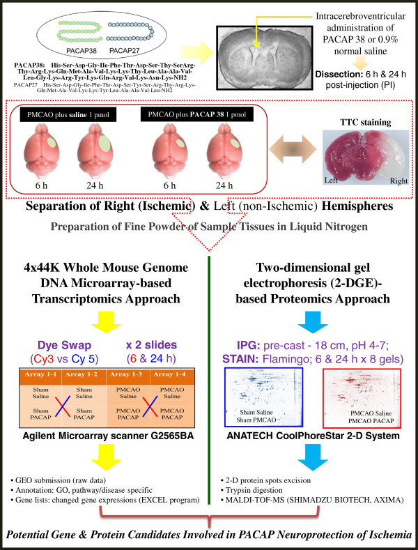
Introduction: The neuropeptide pituitary adenylate cyclase-activating polypeptide (PACAP) is considered to be a potential therapeutic agent for prevention of cerebral ischemia. Ischemia is a most common cause of death after heart attack and cancer causing major negative social and economic consequences. This study was designed to investigate the effect of PACAP38 injection intracerebroventrically in a mouse model of permanent middle cerebral artery occlusion (PMCAO) along with corresponding SHAM control that used 0.9% saline injection.
Methods: Ischemic and non-ischemic brain tissues were sampled at 6 and 24 hours post-treatment. Following behavioral analyses to confirm whether the ischemia has occurred, we investigated the genome-wide changes in gene and protein expression using DNA microarray chip (4x44K, Agilent) and two-dimensional gel electrophoresis (2-DGE) coupled with matrix assisted laser desorption/ionization-time of flight-mass spectrometry (MALDI-TOF-MS), respectively. Western blotting and immunofluorescent staining were also used to further examine the identified protein factor.
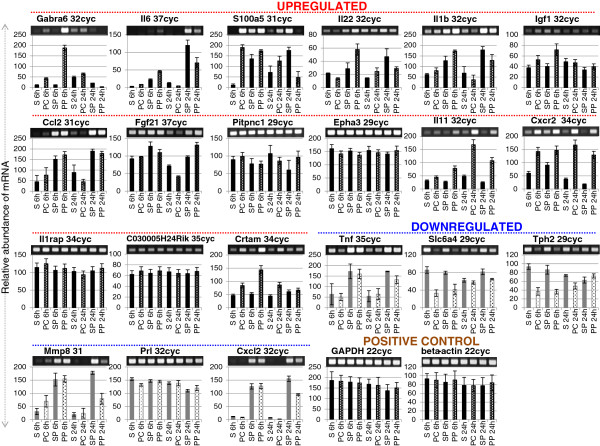
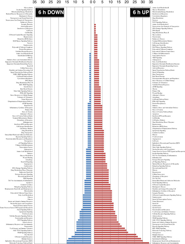
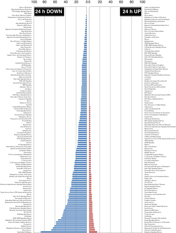
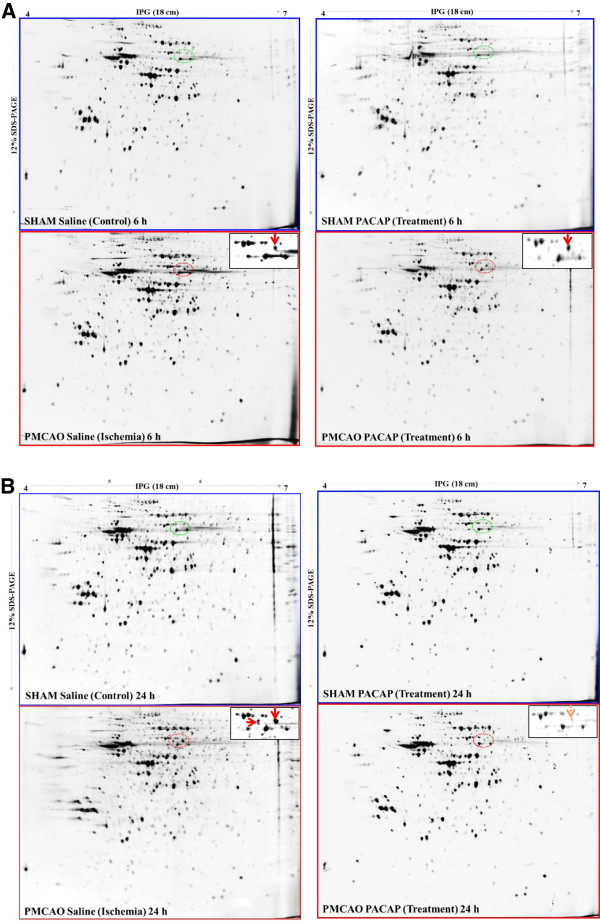
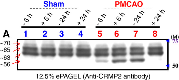
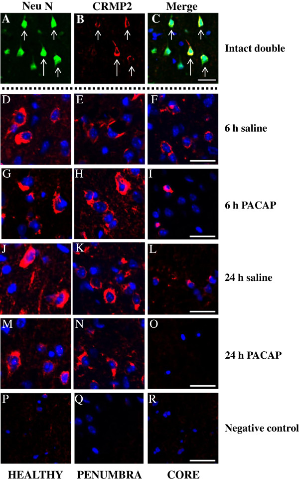
Results: Our results revealed numerous changes in the transcriptome of ischemic hemisphere (ipsilateral) treated with PACAP38 compared to the saline-injected SHAM control hemisphere (contralateral). Previously known (such as the interleukin family) and novel (Gabra6, Crtam) genes were identified under PACAP influence. In parallel, 2-DGE analysis revealed a highly expressed protein spot in the ischemic hemisphere that was identified as dihydropyrimidinase-related protein 2 (DPYL2). The DPYL2, also known as Crmp2, is a marker for the axonal growth and nerve development. Interestingly, PACAP treatment slightly increased its abundance (by 2-DGE and immunostaining) at 6 h but not at 24 h in the ischemic hemisphere, suggesting PACAP activates neuronal defense mechanism early on.
Conclusions: This study provides a detailed inventory of PACAP influenced gene expressions and protein targets in mice ischemic brain, and suggests new targets for thereaupetic interventions.
Figures
Similar articles
-
Molecular Mechanism for PACAP 38-Induced Neurite Outgrowth in PC12 Cells.Neural Plast. 2021 Aug 7;2021:2522454. doi: 10.1155/2021/2522454. eCollection 2021. Neural Plast. 2021. PMID: 34422037 Free PMC article.
-
PACAP38 differentially effects genes and CRMP2 protein expression in ischemic core and penumbra regions of permanent middle cerebral artery occlusion model mice brain.Int J Mol Sci. 2014 Sep 23;15(9):17014-34. doi: 10.3390/ijms150917014. Int J Mol Sci. 2014. PMID: 25257527 Free PMC article.
-
Unraveling the Specific Ischemic Core and Penumbra Transcriptome in the Permanent Middle Cerebral Artery Occlusion Mouse Model Brain Treated with the Neuropeptide PACAP38.Microarrays (Basel). 2015 Jan 21;4(1):2-24. doi: 10.3390/microarrays4010002. Microarrays (Basel). 2015. PMID: 27600210 Free PMC article.
-
PACAP as a neuroprotective factor in ischemic neuronal injuries.Peptides. 2015 Oct;72:202-7. doi: 10.1016/j.peptides.2015.08.006. Epub 2015 Aug 11. Peptides. 2015. PMID: 26275482 Review.
-
Role of PACAP in ischemic neural death.J Mol Neurosci. 2008 Nov;36(1-3):16-25. doi: 10.1007/s12031-008-9077-3. Epub 2008 May 16. J Mol Neurosci. 2008. PMID: 18483879 Review.
Cited by
-
Molecular Mechanism for PACAP 38-Induced Neurite Outgrowth in PC12 Cells.Neural Plast. 2021 Aug 7;2021:2522454. doi: 10.1155/2021/2522454. eCollection 2021. Neural Plast. 2021. PMID: 34422037 Free PMC article.
-
PACAP stimulates functional recovery after spinal cord injury through axonal regeneration.J Mol Neurosci. 2014 Nov;54(3):380-7. doi: 10.1007/s12031-014-0338-z. Epub 2014 Jul 31. J Mol Neurosci. 2014. PMID: 25074795
-
iTRAQ-based proteomic profiling reveals protein alterations after traumatic brain injury and supports thyroxine as a potential treatment.Mol Brain. 2021 Jan 27;14(1):25. doi: 10.1186/s13041-021-00739-0. Mol Brain. 2021. PMID: 33504361 Free PMC article.
-
Bioinformatics investigation of therapeutic mechanisms of Xuesaitong capsule treating ischemic cerebrovascular rat model with comparative transcriptome analysis.Am J Transl Res. 2016 May 15;8(5):2438-49. eCollection 2016. Am J Transl Res. 2016. PMID: 27347353 Free PMC article.
-
PACAP38 differentially effects genes and CRMP2 protein expression in ischemic core and penumbra regions of permanent middle cerebral artery occlusion model mice brain.Int J Mol Sci. 2014 Sep 23;15(9):17014-34. doi: 10.3390/ijms150917014. Int J Mol Sci. 2014. PMID: 25257527 Free PMC article.
References
-
- Miyata A, Jiang L, Dahl RD, Kitada C, Kubo K, Fujino M, Minamino N, Arimura A. Isolation of a neuropeptide corresponding to the N-terminal 27 residues of the pituitary adenylate cyclase activating polypeptide with 38 residues (PACAP38) Biochem Biophys Res Commun. 1990;170:643–648. doi: 10.1016/0006-291X(90)92140-U. - DOI - PubMed
-
- Kimura C, Ohkubo S, Ogi K, Hosoya M, Itoh Y, Onda H, Miyata A, Jian L, Dahl RR, Stibbs HH, Arimura AA, Fujino M. A novel peptide which stimulates adenylate cyclase: molecular cloning and characterization of the ovine and human cDNAs. Biochem Biophys Res Commun. 1990;166:81–89. doi: 10.1016/0006-291X(90)91914-E. - DOI - PubMed
-
- Fahrenkrug J. VIP and PACAP. Results Probl Cell Differ. 2010;50:221–234. - PubMed
Publication types
MeSH terms
Substances
LinkOut - more resources
Full Text Sources
Miscellaneous
