

Inflammatory events during murine squamous cell carcinoma development
- PMID: 23176085
- PMCID: PMC3542019
- DOI: 10.1186/1476-9255-9-46
Inflammatory events during murine squamous cell carcinoma development
Abstract
Background: Squamous cell carcinoma (SCC) is one of the most common human cancers worldwide. In SCC, tumour development is accompanied by an immune response that leads to massive tumour infiltration by inflammatory cells, and consequently, local and systemic production of cytokines, chemokines and other mediators. Studies in both humans and animal models indicate that imbalances in these inflammatory mediators are associated with cancer development.
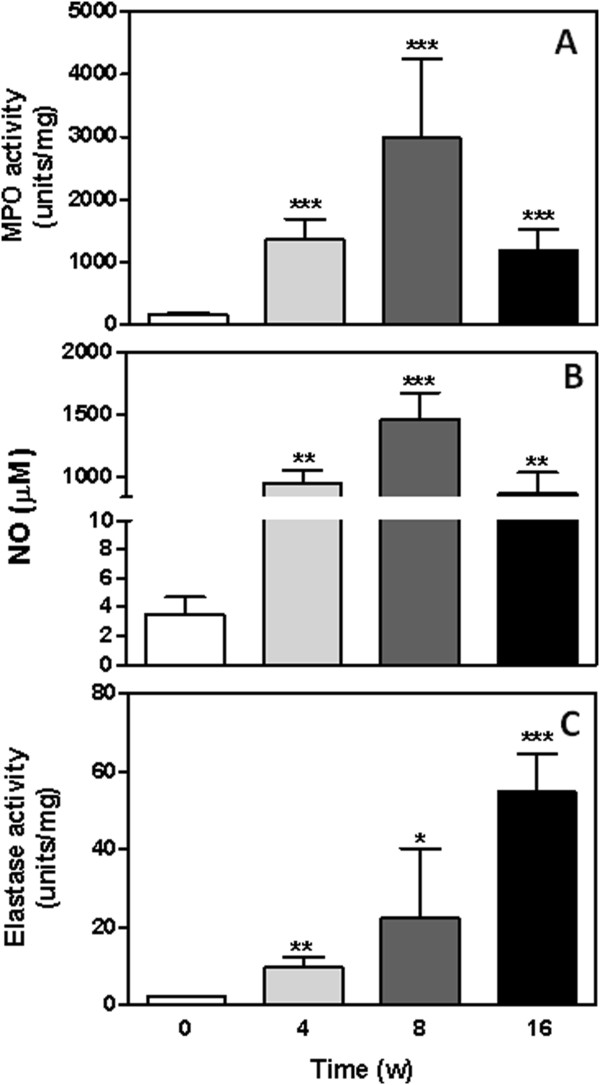
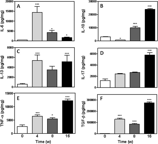
Methods: We used a multistage model of SCC to examine the involvement of elastase (ELA), myeloperoxidase (MPO), nitric oxide (NO), cytokines (IL-6, IL-10, IL-13, IL-17, TGF-β and TNF-α), and neutrophils and macrophages in tumour development. ELA and MPO activity and NO, IL-10, IL -17, TNF-α and TGF-β levels were increased in the precancerous microenvironment.
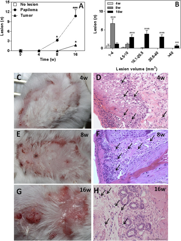
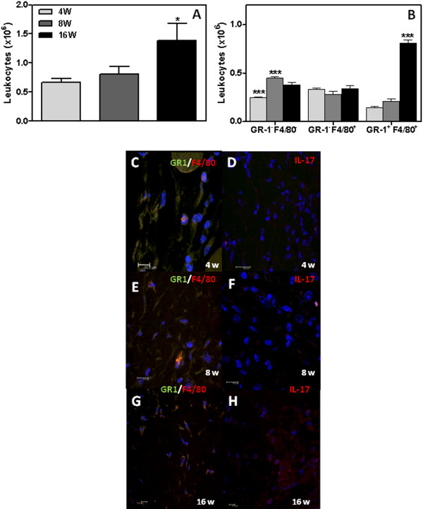
Results: ELA and MPO activity and NO, IL-10, IL -17, TNF-α and TGF-β levels were increased in the precancerous microenvironment. Significantly higher levels of IL-6 and lower levels of IL-10 were detected at 4 weeks following 7,12-Dimethylbenz(a)anthracene (DMBA) treatment. Similar levels of IL-13 were detected in the precancerous microenvironment compared with control tissue. We identified significant increases in the number of GR-1+ neutrophils and F4/80+/GR-1- infiltrating cells in tissues at 4 and 8 weeks following treatment and a higher percentage of tumour-associated macrophages (TAM) expressing both GR-1 and F4/80, an activated phenotype, at 16 weeks. We found a significant correlation between levels of IL-10, IL-17, ELA, and activated TAMs and the lesions. Additionally, neutrophil infiltrate was positively correlated with MPO and NO levels in the lesions.
Conclusion: Our results indicate an imbalance of inflammatory mediators in precancerous SCC caused by neutrophils and macrophages and culminating in pro-tumour local tissue alterations.
Figures
Similar articles
-
Increased elastase and myeloperoxidase activity associated with neutrophil recruitment by IL-17 in airways in vivo.J Allergy Clin Immunol. 2000 Jan;105(1 Pt 1):143-9. doi: 10.1016/s0091-6749(00)90189-1. J Allergy Clin Immunol. 2000. PMID: 10629464
-
Cooperation of liver cells in health and disease.Adv Anat Embryol Cell Biol. 2001;161:III-XIII, 1-151. doi: 10.1007/978-3-642-56553-3. Adv Anat Embryol Cell Biol. 2001. PMID: 11729749 Review.
-
Myeloperoxidase negatively regulates the expression of proinflammatory cytokines and chemokines by zymosan-induced mouse neutrophils.Inflamm Res. 2016 Feb;65(2):151-9. doi: 10.1007/s00011-015-0899-5. Epub 2015 Nov 14. Inflamm Res. 2016. PMID: 26573963
-
Anti-inflammatory and antitumoural effects of Uncaria guianensis bark.J Ethnopharmacol. 2013 Dec 12;150(3):1154-62. doi: 10.1016/j.jep.2013.10.055. Epub 2013 Nov 7. J Ethnopharmacol. 2013. PMID: 24212077
-
Immunological Approaches Towards Cancer and Inflammation: A Cross Talk.Front Immunol. 2018 Mar 20;9:563. doi: 10.3389/fimmu.2018.00563. eCollection 2018. Front Immunol. 2018. PMID: 29662489 Free PMC article. Review.
Cited by
-
Th17 Cells in Protection from Tumor or Promotion of Tumor Progression.J Clin Cell Immunol. 2016 Jun;7(3):431. doi: 10.4172/2155-9899.1000431. Epub 2016 Jun 20. J Clin Cell Immunol. 2016. PMID: 27453801 Free PMC article.
-
Stromal Factors as a Target for Immunotherapy in Melanoma and Non-Melanoma Skin Cancers.Int J Mol Sci. 2022 Apr 6;23(7):4044. doi: 10.3390/ijms23074044. Int J Mol Sci. 2022. PMID: 35409404 Free PMC article. Review.
-
Detection of Squamous Cell Carcinoma in Digitized Histological Images from the Head and Neck Using Convolutional Neural Networks.Proc SPIE Int Soc Opt Eng. 2019 Feb;10956:109560K. doi: 10.1117/12.2512570. Epub 2019 Mar 18. Proc SPIE Int Soc Opt Eng. 2019. PMID: 32476700 Free PMC article.
-
Evaluate the Role of Phosphodiesterase, Myeloperoxidase and Iron in Oral Cancer.Asian Pac J Cancer Prev. 2024 Nov 1;25(11):3921-3926. doi: 10.31557/APJCP.2024.25.11.3921. Asian Pac J Cancer Prev. 2024. PMID: 39611916 Free PMC article.
-
Resistance of R-Ras knockout mice to skin tumour induction.Sci Rep. 2015 Jul 2;5:11663. doi: 10.1038/srep11663. Sci Rep. 2015. PMID: 26133397 Free PMC article.
References
LinkOut - more resources
Full Text Sources
Research Materials
Miscellaneous
