Review
. 2013 Jan;280(2):346-78.
doi: 10.1111/febs.12077. Epub 2013 Jan 17.

Protein tyrosine phosphatases--from housekeeping enzymes to master regulators of signal transduction

Affiliations
Review

Protein tyrosine phosphatases--from housekeeping enzymes to master regulators of signal transduction

Nicholas K Tonks. FEBS J. 2013 Jan.

Abstract

There are many misconceptions surrounding the roles of protein phosphatases in the regulation of signal transduction, perhaps the most damaging of which is the erroneous view that these enzymes exert their effects merely as constitutively active housekeeping enzymes. On the contrary, the phosphatases are critical, specific regulators of signalling in their own right and serve an essential function, in a coordinated manner with the kinases, to determine the response to a physiological stimulus. This review is a personal perspective on the development of our understanding of the protein tyrosine phosphatase family of enzymes. I have discussed various aspects of the structure, regulation and function of the protein tyrosine phosphatase family, which I hope will illustrate the fundamental importance of these enzymes in the control of signal transduction.


Figures

Figure 1
Figure 1. Protein phosphatases in signal transduction
The phosphatases that are implicated in the regulation of signal transduction are highlighted on the left. They are represented by structurally and mechanistically distinct families. The major categories include the PPP and PPM Ser/Thr phosphatases [3, 47], the haloacid dehalogenase (HADS) [226] and the largest group, the Cys-dependent PTP family [44]. The breakdown of the PTPs into individual categories is shown on the right [44].
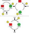
Figure 2
Figure 2. Signaling function of Protein Tyrosine Phosphatases
Members of the PTP family have the potential to act negatively in the regulation of signaling, by dephosphorylating autophosphorylation sites in PTKs themselves or phosphorylation sites in their downstream targets (upper panel). In addition, PTPs may play a positive role, for example by dephosphorylating an inhibitory site in a PTK, such as the C-terminal sites in SRC family PTKs, thereby activating the kinase and promoting phosphorylation and signaling (lower panel).
Figure 3
Figure 3. The sequence motifs that define the conserved PTP catalytic domain
The figure illustrates a ribbon diagram of a PTP catalytic domain, highlighting the positions of the conserved motifs (M1-M10) that define the domain. Areas of conservation (blue = most conserved, red = least conserved) are illustrated using the catalytic domain of PTP1B as the reference. Reproduced from [42], copyright American Society for Microbiology.
Figure 4
Figure 4. Comparison of the active site of different members of the PTP family
A section through the active site of PTP1B (classical, pTyr-specific enzyme), VHR (DUSP) and PTEN (specificity for inositol sugar head group of phosphatidylinositol phospholipid as substrate), to illustrate how the architecture of the active site of members of the PTP family is adapted for their substrate preference (re-drawn from [139]).
Figure 5
Figure 5. Activation of SHP2
In the basal state the active site of SHP2 is occluded by an intramolecular interaction with the N-terminal SH2 domain. The phosphatase may be activated either by engagement of the SH2 domains by pTyr sequence motifs in an RPTK or scaffolding molecule, or by mutations in either the N-SH2 or PTP domains that disrupt their interaction [92].
Figure 6
Figure 6. Mutual regulation of MKP3 and ERK
A Kinase Interaction Motif (KIM) in MKP3 interacts with ERK specifically, inducing a conformational change that promotes MKP function. Phosphorylation of MKP3 by the bound ERK promotes down regulation of MKP by proteolysis [227].
Figure 7
Figure 7. Regulation of the actin cytoskeleton by slingshot phosphatases
Slingshot regulates the actin cytoskeleton through dephosphorylation of cofilin as substrate. The regulation of slingshot illustrates cascade arrangements of both kinases and phosphatases.
Figure 8
Figure 8. KAP1 and regulation of the cell cycle
KAP1 regulates the cell cycle through dephosphorylation of Thr 160 in the activation loop of the cyclin-dependent kinases.
Figure 9
Figure 9. Specific inhibitors of PTP1B
Potent, specific and reversible inhibitors of PTP1B have been generated, both in industry and in academia (illustrated here for the work of ZY Zhang [218]). Unfortunately, many, such as these, are analogues of a pTyr substrate and are thus highly charged, with limited bioavailability.

References

    1. Tonks NK, Diltz CD, Fischer EH. Purification of the major protein tyrosine phosphatases of human placenta. J Biol Chem. 1988;263:6722–6730. - PubMed
    1. Tonks NK, Diltz CD, Fischer EH. Characterization of the major protein tyrosine phosphatases of human placenta. J Biol Chem. 1988;263:6731–6737. - PubMed
    1. Brautigan DL. Protein Ser/ Thr phosphatases - the ugly ducklings of cell signalling. Febs J. 2012 - PubMed
    1. Krebs EG. Protein phosphorylation and cellular regulation I. Nobel Lecture. 1992:72–89. - PubMed
    1. Fischer EH. Protein phosphorylation and cellular regulation II. Nobel Lecture. 1992:95–113.

Publication types

MeSH terms

Substances