

Smart surface for elution of protein-protein bound particles: nanonewton dielectrophoretic forces using atomic layer deposited oxides
- PMID: 23176521
- PMCID: PMC4984534
- DOI: 10.1021/ac302857z
Smart surface for elution of protein-protein bound particles: nanonewton dielectrophoretic forces using atomic layer deposited oxides
Abstract
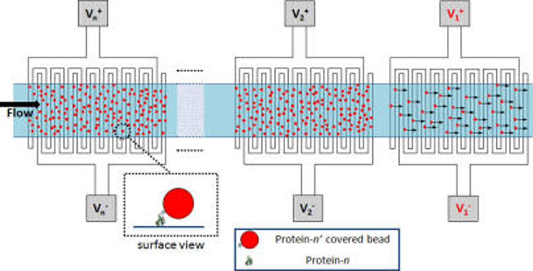
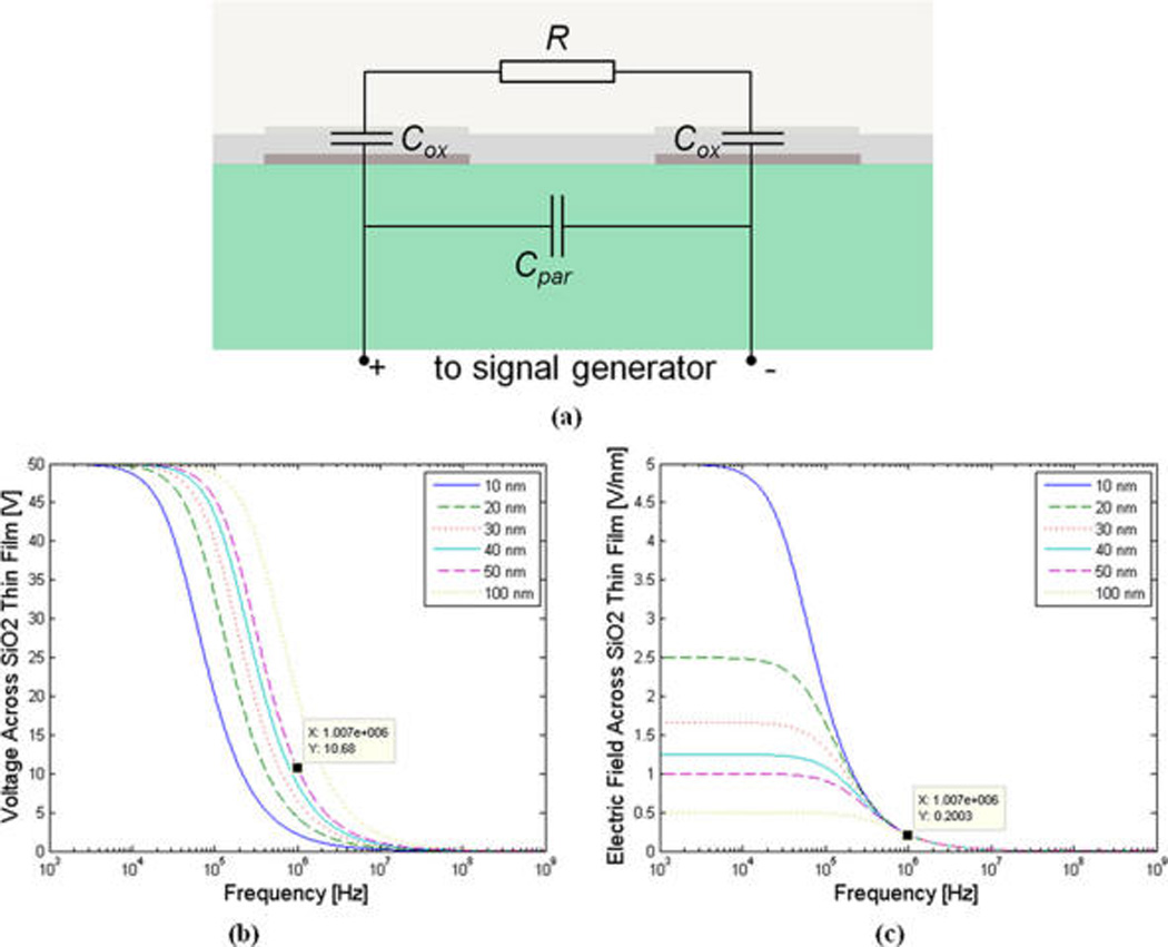
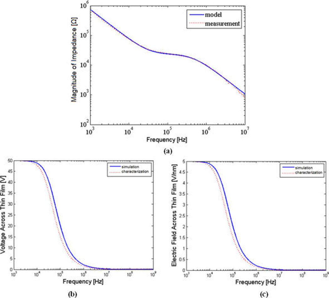
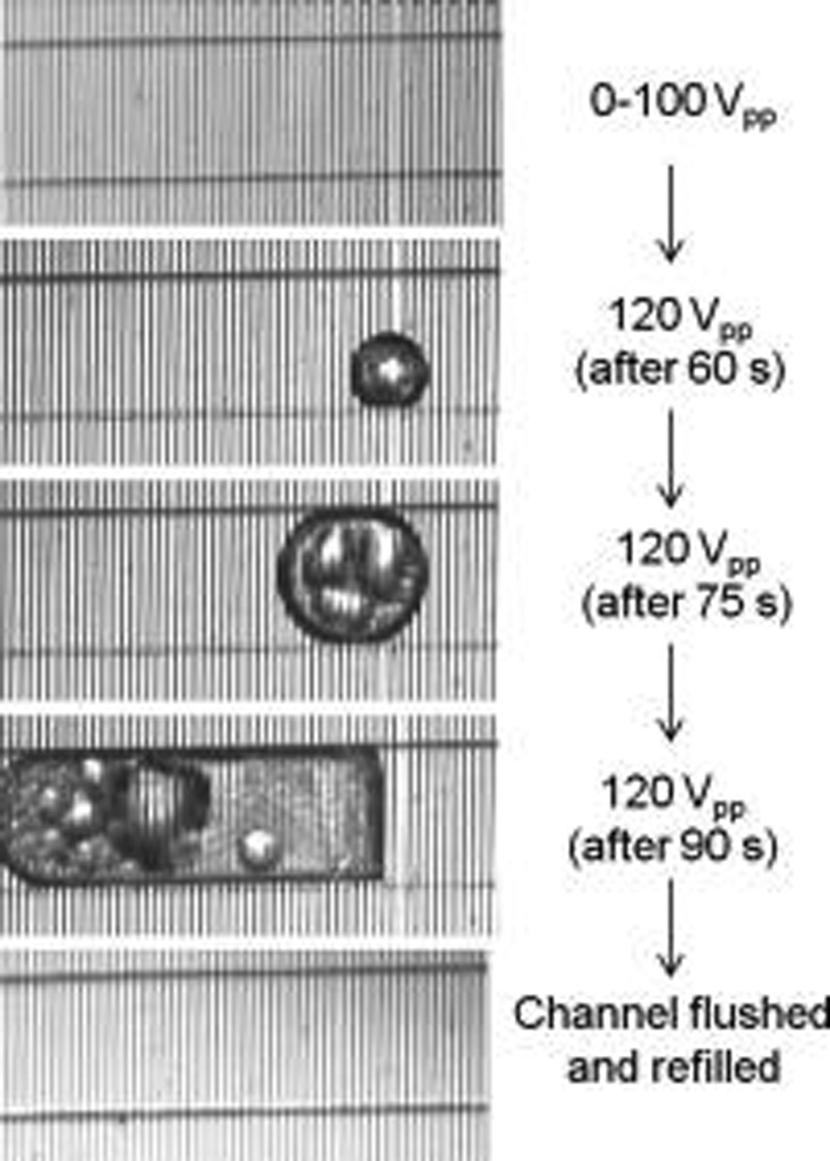
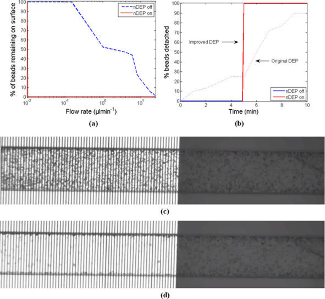
By increasing the strength of the negative dielectrophoresis force, we demonstrated a significantly improved electrokinetic actuation and switching microsystem that can be used to elute specifically bound beads from the surface. In this work using atomic layer deposition we deposited a pinhole free nanometer-scale thin film oxide as a protective layer to prevent electrodes from corrosion, when applying high voltages (>20 V(pp)) at the electrodes. Then, by exciting the electrodes at high frequency, we capacitively coupled the electrodes to the buffer in order to avoid electric field degradation and, hence, reduction in dielectrophoresis force due to the presence of the insulating oxide layer. To illustrate the functionality of our system, we demonstrated 100% detachment of anti-IgG and IgG bound beads (which is on the same order of magnitude in strength as typical antibody-antigen interactions) from the surface, upon applying the improved negative dielectrophoresis force. The significantly enhanced switching performance presented in this work shows orders of magnitude of improvement in on-to-off ratio and switching response time, without any need for chemical eluting agents, as compared to the previous work. The promising results from this work vindicates that the functionality of this singleplexed platform can be extended to perform a multiplexed bead-based assay where in a single channel an array of proteins are patterned each targeting a different antigen or protein.
Figures
Similar articles
-
Multiplexed actuation using ultra dielectrophoresis for proteomics applications: a comprehensive electrical and electrothermal design methodology.Lab Chip. 2014 Jun 21;14(12):2105-14. doi: 10.1039/c4lc00036f. Epub 2014 May 7. Lab Chip. 2014. PMID: 24801800 Free PMC article.
-
Use of negative dielectrophoresis for selective elution of protein-bound particles.Anal Chem. 2012 Feb 7;84(3):1432-8. doi: 10.1021/ac202508u. Epub 2012 Jan 20. Anal Chem. 2012. PMID: 22242790 Free PMC article.
-
Dielectrophoresis in microchips containing arrays of insulating posts: theoretical and experimental results.Anal Chem. 2003 Sep 15;75(18):4724-31. doi: 10.1021/ac0340612. Anal Chem. 2003. PMID: 14674447
-
Electric field-induced effects on neuronal cell biology accompanying dielectrophoretic trapping.Adv Anat Embryol Cell Biol. 2003;173:III-IX, 1-77. doi: 10.1007/978-3-642-55469-8. Adv Anat Embryol Cell Biol. 2003. PMID: 12901336 Review.
-
Experimental study of dielectrophoresis and liquid dielectrophoresis mechanisms for particle capture in a droplet.Electrophoresis. 2011 Jun;32(11):1337-47. doi: 10.1002/elps.201000548. Epub 2011 Apr 28. Electrophoresis. 2011. PMID: 21538398
Cited by
-
A 96-wells fluidic system for high-throughput screenings under laminar high wall shear stress conditions.Microsyst Nanoeng. 2023 Sep 15;9:114. doi: 10.1038/s41378-023-00589-x. eCollection 2023. Microsyst Nanoeng. 2023. PMID: 37719414 Free PMC article.
-
High Sensitivity Extended Nano-Coulter Counter for Detection of Viral Particles and Extracellular Vesicles.Anal Chem. 2023 Jul 4;95(26):9892-9900. doi: 10.1021/acs.analchem.3c00855. Epub 2023 Jun 19. Anal Chem. 2023. PMID: 37336762 Free PMC article.
-
Portable Cytometry Using Microscale Electronic Sensing.Sens Actuators B Chem. 2016 Mar 1;224:275-281. doi: 10.1016/j.snb.2015.08.118. Epub 2015 Sep 2. Sens Actuators B Chem. 2016. PMID: 27647950 Free PMC article.
-
Microfluidic and Nanofluidic Resistive Pulse Sensing: A Review.Micromachines (Basel). 2017 Jun 25;8(7):204. doi: 10.3390/mi8070204. Micromachines (Basel). 2017. PMID: 30400393 Free PMC article. Review.
-
Study on non-bioparticles and Staphylococcus aureus by dielectrophoresis.RSC Adv. 2020 Jan 15;10(5):2598-2614. doi: 10.1039/c9ra05886a. eCollection 2020 Jan 14. RSC Adv. 2020. PMID: 35496126 Free PMC article.
References
-
- Hamburg MA, Collins FS. N. Engl. J. Med. 2010;363(4):301–304. - PubMed
-
- Harvey A, Brand A, Holgate ST, Kristiansen LV, Lehrach H, Palotie A, Prainsack B. New Biotechnol. 2012;29(6):625–633. - PubMed
-
- Wei DQ, Chen Q. Curr. Drug Metab. 2012;13(7):951–951. - PubMed
-
- Souied EH, Leveziel N. Am. J. Ophthalmol. 2012;154(3):427–428. - PubMed
Publication types
MeSH terms
Substances
Grants and funding
LinkOut - more resources
Full Text Sources
Miscellaneous
