

A longitudinal analysis of risk factors associated with central retinal vein occlusion
- PMID: 23177364
- PMCID: PMC3563864
- DOI: 10.1016/j.ophtha.2012.07.080
A longitudinal analysis of risk factors associated with central retinal vein occlusion
Abstract
Purpose: To identify risk factors associated with central retinal vein occlusion (CRVO) among a diverse group of patients throughout the United States.
Design: Longitudinal cohort study.
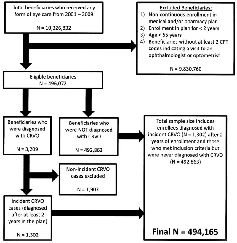
Participants: All beneficiaries aged ≥ 55 years who were continuously enrolled in a managed care network for at least 2 years and who had ≥ 2 visits to an eye care provider from 2001 to 2009.
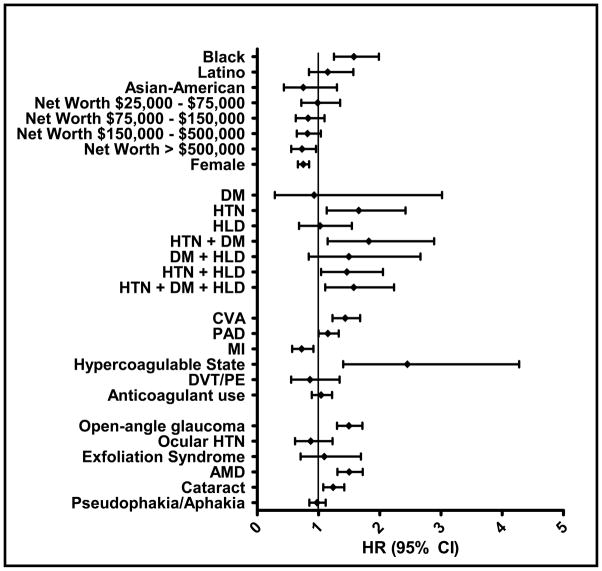
Methods: Insurance billing codes were used to identify individuals with a newly diagnosed CRVO. Multivariable Cox regression was performed to determine the factors associated with CRVO development.
Main outcome measures: Adjusted hazard ratios (HRs) with 95% confidence intervals (CIs) of being diagnosed with CRVO.
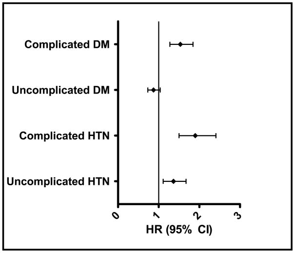
Results: Of the 494 165 enrollees who met the study inclusion criteria, 1302 (0.26%) were diagnosed with CRVO over 5.4 (± 1.8) years. After adjustment for known confounders, blacks had a 58% increased risk of CRVO compared with whites (HR, 1.58; 95% CI, 1.25-1.99), and women had a 25% decreased risk of CRVO compared with men (HR, 0.75; 95% CI, 0.66-0.85). A diagnosis of stroke increased the hazard of CRVO by 44% (HR, 1.44; 95% CI, 1.23-1.68), and hypercoagulable state was associated with a 145% increased CRVO risk (HR, 2.45; 95% CI, 1.40-4.28). Individuals with end-organ damage from hypertension (HTN) or diabetes mellitus (DM) had a 92% (HR, 1.92; 95% CI, 1.52-2.42) and 53% (HR, 1.53; 95% CI, 1.28-1.84) increased risk of CRVO, respectively, relative to those without these conditions.
Conclusions: This study confirms that HTN and vascular diseases are important risk factors for CRVO. We also identify black race as being associated with CRVO, which was not well appreciated previously. Furthermore, we show that compared with patients without DM, individuals with end-organ damage from DM have a heightened risk of CRVO, whereas those with uncomplicated DM are not at increased risk of CRVO. This finding may provide a potential explanation for the conflicting reports in the literature on the association between CRVO and DM. Information from analyses such as this can be used to create a risk calculator to identify possible individuals at greatest risk for CRVO.
Copyright © 2013 American Academy of Ophthalmology. Published by Elsevier Inc. All rights reserved.
Conflict of interest statement
No conflicting relationship exists for any author regarding any material discussed in this manuscript.
Figures
References
-
- Cugati S, Wang JJ, Rochtchina E, Mitchell P. Ten-year incidence of retinal vein occlusion in an older population: the Blue Mountains Eye Study. Arch Ophthalmol. 2006;124:726–32. - PubMed
-
- Branch Vein Occlusion Study Group. Argon laser photocoagulation for macular edema in branch vein occlusion. Am J Ophthalmol. 1984;98:271–82. - PubMed
-
- Central Vein Occlusion Study Group. Natural history and clinical management of central retinal vein occlusion. Arch Ophthalmol. 1997;115:486–91. - PubMed
Publication types
MeSH terms
Grants and funding
LinkOut - more resources
Full Text Sources
Other Literature Sources
Medical
Miscellaneous
