. 2012 Nov 21;76(4):838-846.
doi: 10.1016/j.neuron.2012.09.029.

Synchronous oscillatory neural ensembles for rules in the prefrontal cortex

Affiliations

Synchronous oscillatory neural ensembles for rules in the prefrontal cortex

Timothy J Buschman et al. Neuron. .

Abstract

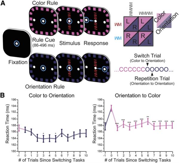
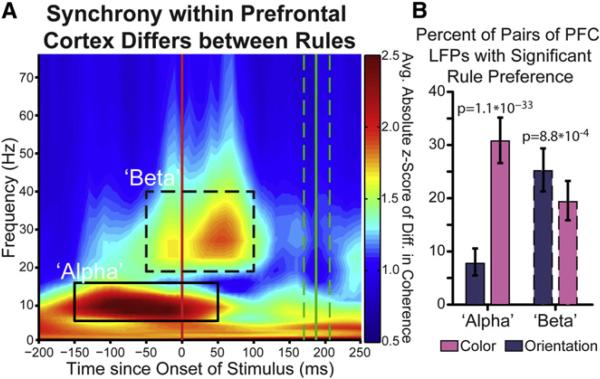
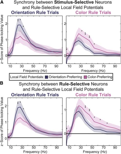
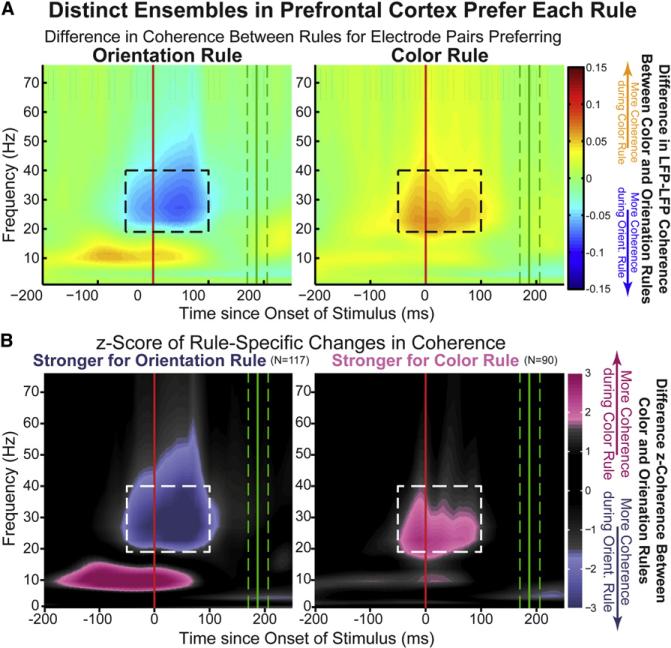
Intelligent behavior requires acquiring and following rules. Rules define how our behavior should fit different situations. To understand its neural mechanisms, we simultaneously recorded from multiple electrodes in dorsolateral prefrontal cortex (PFC) while monkeys switched between two rules (respond to color versus orientation). We found evidence that oscillatory synchronization of local field potentials (LFPs) formed neural ensembles representing the rules: there were rule-specific increases in synchrony at "beta" (19-40 Hz) frequencies between electrodes. In addition, individual PFC neurons synchronized to the LFP ensemble corresponding to the current rule (color versus orientation). Furthermore, the ensemble encoding the behaviorally dominant orientation rule showed increased "alpha" (6-16 Hz) synchrony when preparing to apply the alternative (weaker) color rule. This suggests that beta-frequency synchrony selects the relevant rule ensemble, while alpha-frequency synchrony deselects a stronger, but currently irrelevant, ensemble. Synchrony may act to dynamically shape task-relevant neural ensembles out of larger, overlapping circuits.


Figures

Figure 1
Figure 1. Task Design and Behavioral Performance
(A) Task timeline. Eye position is indicated by the blue circles. Animals initiated trial by fixating the center dot. After presentation of a border cue indicating the rule, the stimulus was presented. The animal integrated the rule and stimulus in order to make a decision about the required saccade: under the color rule, red stimuli meant saccade left and blue stimuli meant saccade right; under the orientation rule, vertical meant saccade right and horizontal meant saccade left. The rule in effect was blocked and switched randomly after a minimum of 20 trials. (B) An asymmetric cost was observed when switching between rules, reflected in the speed at which the animals performed the task. Switching from orientation to color was significantly slower, but no cost was observed when switching from color to orientation. This suggests that orientation was behaviorally dominant. All error bars represent SEM. ***p ≤ 10−3; **p ≤ 0.01; *p ≤ 0.05.
Figure 2
Figure 2. PFC Neurons Encode Task-Relevant Information Including the Current Rule and Stimulus
(A) Information about the current rule (black line) is captured using a bias-corrected percent explained variance statistic (y axis) and is determined in a sliding window across the trial (x axis). Shaded region indicates 95% confidence interval. As the rule often repeated on consecutive trials (see Figure 1A), there was some expectancy of the rule encoded by PFC neurons before rule cue onset (although not significant across the population of recorded PFC neurons). (B) PFC neurons encode stimulus identity, both its orientation (green line) and color (blue line). Shaded regions indicate 95% confidence interval. Information about the orientation of the stimulus was more strongly represented across the population, possibly leading to the behavioral dominance of the orientation rule (see Figure 1B).
Figure 3
Figure 3. Rule-Selective Synchrony in PFC
(A) Synchrony between electrodes within prefrontal cortex differs for rules. Synchrony is quantified by the coherence in simultaneously recorded local field potentials during each rule. The difference in synchrony (rectified to capture synchrony differences that prefer either rule) was compared to a trial-shuffled null distribution, resulting in a Z score of observed rule difference (color axis). Absolute synchrony differences are shown across time relative to stimulus onset (x axis) and frequency (y axis). Two time-frequency regions of interest (ROIs) are seen—an “alpha,” 6–16 Hz, prestimulus ROI (solid outline) and a “beta,” 19–40 Hz, peristimulus ROI (dashed outline). (B) Percentage of recorded pairs of electrodes with a significant rule preference during the “alpha” and “beta” time-frequency regions of interest (solid/ dashed outlines in A). Error bars indicate 95% confidence interval. Significantly more electrode pairs prefer color within the alpha ROI and orientation within the beta ROI.
Figure 4
Figure 4. Magnitude of Rule-Selective Changes in Synchrony
(A) Individual electrode pairs in the beta ROI are highly synchronous and show significant rule-dependent change. Coherence between rule-dependent pairs of electrodes (pink and purple +, main panel; group averages, solid circles) in the beta ROI was high overall (cumulative probability distribution, bottom) and generally reflected a 10% or greater change in coherence over the non-preferred rule (histogram, right) compared to no-rule-preferring electrode pairs (gray x, main panel). (B) Average difference in coherence between preferred and nonpreferred rules for all beta ROI electrode pairs.
Figure 5
Figure 5. Single Neurons Carrying Task-Relevant Information Synchronize to the Currently Relevant Ensemble
Neurons encoding task-relevant information were more synchronized with the rule-selective ensemble preferring the current rule. Phase locking of stimulus-selective neurons (A) and rule-selective neurons (B) to electrodes that participated in either the color-preferring ensemble (pink) or the orientation-preferring ensemble (purple). Only electrodes that were exclusive to either ensemble were used (i.e., those electrodes participating in both ensembles were excluded). Phase locking is shown for both orientation trials (left) and color trials (right). Shaded regions indicate 95% confidence intervals. Significant differences in phase locking between the two ensembles are indicated at each frequency tested (*p < 0.05; **p < 0.01).
Figure 6
Figure 6. Independent, Rule-Specific PFC Ensembles
Ensembles within PFC can be identified by rule-selective synchrony in the peristimulus “beta” ROI (dashed outline). One ensemble is more synchronous during orientation trials (A, left). This difference is significantly greater than expected by chance (B, left). A separate ensemble of electrodes is more synchronous during color trials (A, right). Again, this difference is significant (B, right). Alpha-band synchrony is observed in the orientation ensemble during the competing color rule (left panels, orange and pink colors for top and bottom rows) but not in the color ensemble (right) or during the orientation rule (Figure 2B). Axes are the same as Figure 3A, but now color axes are no longer rectified: orange (top row) and pink (bottom row) reflect greater synchrony during color rule trials; blue (top row) and purple (bottom row) during orientation rule trials. Please note the color axis of (B) is intentionally nonlinear, showing only significant rule selectivity, beginning at a Z score of ±1.67 (p = 0.05) and fully saturated at ±1.97 (p = 0.01).
Figure 7
Figure 7. Strength of Prefrontal Synchrony Selectivity Correlates with Reaction Time
Trials in which the monkeys responded faster (left) showed stronger rule-selective synchrony in the “alpha” and “beta” regions of interest compared to trials with slower reaction times (right). Green lines indicate reaction time quartiles and white lines indicate the corresponding preparatory period quartiles. Black lines on faster reaction time trials (left) indicate when synchrony in the alpha-and beta-frequency bands (gray and black diamonds, respectively) was significantly higher than synchrony during slower-reaction time trials.

Comment in

References

    1. Aertsen AM, Gerstein GL, Habib MK, Palm G. Dynamics of neuronal firing correlation: modulation of “effective connectivity”. J. Neurophysiol. 1989;61:900–917. - PubMed
    1. Akam T, Kullmann DM. Oscillations and filtering networks support flexible routing of information. Neuron. 2010;67:308–320. - PMC - PubMed
    1. Allport DA, Styles EA, Hsieh S. Shifting intentional set: Exploring the dynamic control of tasks. In: Umilta C, Moscovitch M, editors. Attention and Performance XV: Conscious and Nonconscious Information Processing. The MIT Press; Cambridge, MA: 1994. pp. 421–452.
    1. Androulidakis AG, Doyle LMF, Yarrow K, Litvak V, Gilbertson TP, Brown P. Anticipatory changes in beta synchrony in the human corticospinal system and associated improvements in task performance. Eur. J. Neurosci. 2007;25:3758–3765. - PubMed
    1. Axmacher N, Schmitz DP, Wagner T, Elger CE, Fell J. Interactions between medial temporal lobe, prefrontal cortex, and inferior temporal regions during visual working memory: a combined intracranial EEG and functional magnetic resonance imaging study. J. Neurosci. 2008;28:7304–7312. - PMC - PubMed

Publication types

LinkOut - more resources