. 2012 Nov 21;151(5):1083-96.
doi: 10.1016/j.cell.2012.10.023.

Endocardial cells form the coronary arteries by angiogenesis through myocardial-endocardial VEGF signaling

Affiliations

Endocardial cells form the coronary arteries by angiogenesis through myocardial-endocardial VEGF signaling

Bingruo Wu et al. Cell. .

Abstract

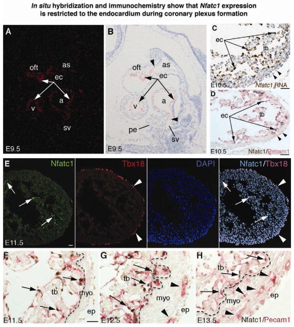
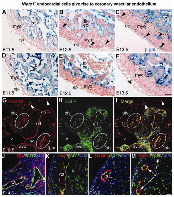
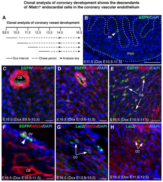
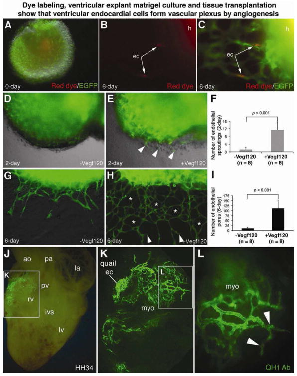
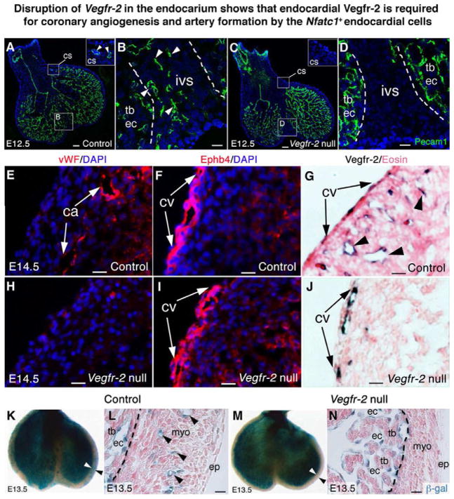
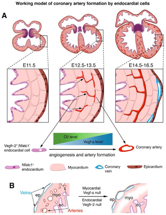
The origins and developmental mechanisms of coronary arteries are incompletely understood. We show here by fate mapping, clonal analysis, and immunohistochemistry that endocardial cells generate the endothelium of coronary arteries. Dye tracking, live imaging, and tissue transplantation also revealed that ventricular endocardial cells are not terminally differentiated; instead, they are angiogenic and form coronary endothelial networks. Myocardial Vegf-a or endocardial Vegfr-2 deletion inhibited coronary angiogenesis and arterial formation by ventricular endocardial cells. In contrast, lineage and knockout studies showed that endocardial cells make a small contribution to the coronary veins, the formation of which is independent of myocardial-to-endocardial Vegf signaling. Thus, contrary to the current view of a common source for the coronary vessels, our findings indicate that the coronary arteries and veins have distinct origins and are formed by different mechanisms. This information may help develop better cell therapies for coronary artery disease.


Figures

Figure 1
Figure 1. In situ hybridization and immunochemistry show that Nfatc1 expression is restricted to the endocardium during coronary plexus formation
(A,B) E9.5 heart sections show Nfatc1 transcripts in the endocardium (ec, arrows). Nfatc1 transcript signals separate the positive endocardium from the negative endothelium of aortic sac (as) and sinus venosus (sv) (arrowheads). Background signals are seen in the proepicardium (pe). a, atrium; oft, outflow tract; v, ventricle (C) E10.5 ventricular sections show Nfatc1 transcripts in the endocardium (arrows) but not epicardium (arrowheads). tb, trabeculae (D) E10.5 heart sections co-immunostained with antibodies against Nfatc1 (brown nuclear staining) and Pecam1 (red membrane staining) show Nfatc1 proteins in the endocardium (arrows) but not epicardium (arrowheads). (E) E11.5 heart sections show Nfatc1 proteins in the endocardium (arrows) but not in the Tbx18-positive epicardium (arrowheads). (FH) E11.5–E13.5 heart sections stained with antibodies against Nfatc1 and Pecam1 show that Nfatc1 proteins are restricted to the endocardium (arrows). They are not present in the endothelium of coronary plexuses or Pecam1-positive cells in the myocardium (myo, arrowheads) or the mature coronary vessels (see also Figure S1). Dashed line separates the trabeculae from the compact wall. ep, epicardium. All bars = 25 μm.
Figure 2
Figure 2. Fate-mapping analysis reveals that Nfatc1+ endocardial cells generate coronary vascular endothelium
(AC) X-gal stained E11.5–E13.5 heart sections of Nfatc1Cre;R26fslz embryos show that the β-gal tagged Nfatc1+ endocardial cells reside in the endocardium of the myocardial wall and trabeculae at E11.5 (A), they begin to invade the myocardium at E12.5 (B, arrowheads) and generate networks of coronary plexuses at E13.5 (C, arrowheads). The descendants of Nfatc1+ cells are not present in the epicardium and myocardium (see also Figures S2 and S3). (DF) X-gal stained E11.5–E13.5 heart sections of control Nfatc1lacZ-BAC embryos show that the β-gal activities directed by the Nfatc1 promoter/enhancer are restricted to the endocardium and not present in the myocardium and epicardium. Unlike Nfatc1Cre, Nfatc1lacZ does not label the coronary plexuses. (GI) Dual fluorescent sections through ventricular (v) outflow tract (oft) of E12.5 Nfatc1Cre;RCEfsEGFP embryos show the EGFP+ endothelial descendants of Nfatc1+ endocardial cells in the Pecam1+ coronary plexuses in the peritruncal region (ptv). The peripheral vessels (arrowheads) expressing Pecam1 but not EGFP are not derived from the Nfatc1+ endocardial cells (see also Figure S4). Mesenchyme of atrioventricular canal (avc, asterisk), derived from the cushion endocardial cells, is EGFP positive but Pecam1 negative. (JK) Dual fluorescent sections of E14.5 Nfatc1Cre;RCEfsEGFP heart show EGFP+ endothelial descendants of endocardial cells in the Dll4+ or vWF+ endothelium (red) of the main coronary arteries (ca). The trabecular endocardium is negative for vWF. (LM) Dual fluorescent sections of E16.5 Nfatc1Cre;RCEfsEGFP heart show EGFP+ endothelial descendants of endocardial cells in the inner layer of the coronary arteries (ca) surrounded by smooth muscle cells positive for SM-MHC or SM22a (see also Figure S5 and S6). All Bars = 25 μm.
Figure 3
Figure 3. Clonal analysis of coronary development shows the descendants of Nfatc1+ endocardial cells in the coronary vascular endothelium
(A) A schematic diagram showing clonal analysis of coronary development using the Nfatc1nrtTA;tetO-Cre system induced by doxycycline (Dox). (B) Heart section of Nfatc1nrtTA;tetO-Cre;RCEfsEGFP E11.5 embryo induced at E10.5 with a limited Dox dose shows single EGFP-labeled cells in the endocardium (arrowhead) (see also Figure S7). (CE) Sections of E16.5 Nfatc1nrtTA;tetO-Cre;RCEfsEGFP heart induced at E9.5 (C) or E10.5 (D,E) show the EGFP+ cell clusters in the large (C, D) or small coronary arteries (arrows) expressing SM22a. (F) Sections of E16.5 Nfatc1nrtTA;tetO-Cre;RCEfsEGFP heart induced at E10.5 shows the EGFP+ cell clusters (arrowhead) in the subepicardial vessels alongside the SM22a+ cells. (G) Sections of E16.5 Nfatc1nrtTA;tetO-Cre;R26fslz heart induced at E12.5 shows the LacZ-labeled capillary cells (cc) cluster (green; arrows) alongside the SM22a-expressing small arteries (red). (H) Sections of E16.5 Nfatc1nrtTA;tetO-Cre;R26fslz heart induced at E11.5 shows the LacZ+ capillary cells cluster (arrows) alongside the Pecam1+ small vessels (red). See also Figures S8, S9, and Table S2. All bars = 20 μm.
Figure 4
Figure 4. Dye labeling, ventricular explant culture and tissue transplantation show that ventricular endocardial cells form the vascular plexus by angiogenesis
(A–C) Dye labeling and explant matrigel cultures (see also Figure S10, A to C) using ventricles from E11.5 Nfatc1Cre;RCEfsEGFP hearts (h) show red fluorescent CMTPX-labeled individual endocardial cells (arrows) that had migrated through the ventricular wall and integrated into an EGFP+ endothelial network upon Vegf120 treatment. (DI) Matrigel angiogenesis assays show that Vegf120 significantly promotes transmural migration, sprouting (arrowheads), and endothelial networking (asterisks) by Nfatc1+ descendants. The error bars represent SD. See also Movies S1 and S2. (JL) Images of QH1 antibody stained HH34 quail-chick chimerical heart sliced through the right ventricle at the implanted site show that the QH1+ descendants of engrafted HH15 quail endocardial cells invade chick myocardium and generate intramyocardial vessels. Quail endocardial cells, implanted at the surface of the atrioventricular junction of the chick heart (K), generate intensive myocardial vascular networks by angiogenic sprouting and branching (L, arrowheads). See also Figure S10, D–F, and Table S3.
Figure 5
Figure 5. Disruption of Vegf-a in the myocardium reveals that myocardial Vegf-a is required for coronary angiogenesis and artery formation
(AD) Pictures of E12.5 Tnnt2Cre;Vegf-a+/+ (Control) heart stained with Pecam1 antibodies exhibit coronary plexuses (arrows) in the peritruncal region and interventricular septum (ivs). Arrowheads indicate the branching endothelial tubes. Dashed lines separate the Pecam1+ vessels in the septum from the Pecam1+ trabeculae. (EH) Pictures of E12.5 Tnnt2Cre;Vegf-af/f (Vegf-a null) heart show no plexus formation in the peritruncal and septal myocardium. (IK) Pictures of E14.5 control heart show the Pecam1+ coronary arteries (ca, arrows) and subepicardial vessels (arrowheads). (LN) Pictures of E14.5 Vegf-a null heart show less and immaturely formed coronary arteries (arrows) but numerous and dilated subepicardial vessels (arrowheads). The null heart also has necrotic peritruncal and septal myocardium (L, arrows) (see also Figures S11, S12, and Table S4). Bar in I,L = 100 μm; rest = 20 μm.
Fig. 6
Fig. 6. Disruption of Vegfr-2 in the endocardium shows that endocardial Vegfr-2 is required for coronary angiogenesis and artery formation by endocardial cells
(A,B) E12.5 Nfatc1Cre;Vegfr-2+/+ (Control) heart sections stained with Pecam1 antibodies show coronary endothelial tubes in the myocardium of the coronary sulcus (cs) (A, inset) and interventricular septum (B, arrowheads). (C,D) E12.5 Nfatc1Cre;Vegfr-2f/f (Vegfr-2 null) heart sections stained with Pecam1 antibodies show no coronary endothelial tubes in the coronary sulcus (C, inset) and interventricular septum (D). Dashed lines separate the septum from the Pecam1+ endocardium of trabeculae. (EG) E14.5 control heart sections show that mature coronary arteries are positive for vWF staining (E, arrows) while subepicardial coronary veins (cv) are positive for Ephb4 staining (F, arrows). Vegfr-2 proteins are present in arteries (G, arrowheads) and veins (G, arrows). (HJ) E14.5 Vegfr-2 null heart sections show no vWF+ coronary arteries in the myocardium (H), but presence of Ephb4+ subepicardial coronary veins (I, arrows). Vegfr-2 proteins are only present in the coronary veins (J, arrows). (K,L) X-gal stained E13.5 Nfatc1Cre;Vegfr-2+/+;R26fslz (Control) heart show that the β-gal+ descendants of Nfatc1+ endocardial cells generate the coronary vessels in the ventricular wall (arrowheads). Dashed lines distinguish the β-gal+ coronary plexuses in the compact wall from the β-gal+ endocardium of trabeculae. (M,N) X-gal stained E13.5 Nfatc1Cre;Vegfr-2f/f;R26fslz (Vegfr-2 null) heart show no β-gal+ coronary plexuses derived from endocardial cells in the ventricular wall. See also Figures S14, S15, and Table S5. Bar in A,C = 100 μm; rest = 20 μm.
Figure 7
Figure 7. Working model of coronary artery formation by endocardial cells
(A) Diagram shows an ontogenic role for endocardial cells in generation of the coronary arteries. Between E11.5 and E13.5 of mouse embryogenesis, myocardial proliferation generates a Vegf-a gradient across the thickening ventricular wall, possibly regulated by a reverse gradient of myocardial O2 content. At the same time, some ventricular endocardial cells turn off nuclear Nfatc1 expression (purple nuclei). The Vegf-a gradient induces these Nfatc1 endocardial cells (white nuclei) to invade the myocardium and proliferate into coronary plexuses by angiogenesis via their expression of Vegfr-2. The plexuses then develop into coronary arteries. (B) Knockout of Vegf-a in the myocardium or Vegfr-2 in the endocardium prevents coronary angiogenesis and artery formation, but does not block coronary vein formation, suggesting that coronary veins arise from non-endocardial origins, independent of myocardial Vegf-a to endocardial Vegfr-2 signaling.

Comment in

References

    1. Cai CL, Martin JC, Sun Y, Cui L, Wang L, Ouyang K, Yang L, Bu L, Liang X, Zhang X, et al. A myocardial lineage derives from Tbx18 epicardial cells. Nature. 2008;454:104–108. - PMC - PubMed
    1. Carmeliet P, Ferreira V, Breier G, Pollefeyt S, Kieckens L, Gertsenstein M, Fahrig M, Vandenhoeck A, Harpal K, Eberhardt C, et al. Abnormal blood vessel development and lethality in embryos lacking a single VEGF allele. Nature. 1996;380:435–439. - PubMed
    1. Chang CP, Neilson JR, Bayle JH, Gestwicki JE, Kuo A, Stankunas K, Graef IA, Crabtree GR. A field of myocardial-endocardial NFAT signaling underlies heart valve morphogenesis.[see comment] Cell. 2004;118:649–663. - PubMed
    1. Chen JW, Zhou B, Yu QC, Shin SJ, Jiao K, Schneider MD, Baldwin HS, Bergelson JM. Cardiomyocyte-specific deletion of the coxsackievirus and adenovirus receptor results in hyperplasia of the embryonic left ventricle and abnormalities of sinuatrial valves. Circ Res. 2006;98:923–930. - PubMed
    1. de la Pompa JL, Timmerman LA, Takimoto H, Yoshida H, Elia AJ, Samper E, Potter J, Wakeham A, Marengere L, Langille BL, et al. Role of the NF-ATc transcription factor in morphogenesis of cardiac valves and septum.[see comment] Nature. 1998;392:182–186. - PubMed

Publication types

MeSH terms

Substances