. 2012 Nov 21;151(5):1113-25.
doi: 10.1016/j.cell.2012.10.024.

A fructose receptor functions as a nutrient sensor in the Drosophila brain

Affiliations

A fructose receptor functions as a nutrient sensor in the Drosophila brain

Tetsuya Miyamoto et al. Cell. .

Abstract

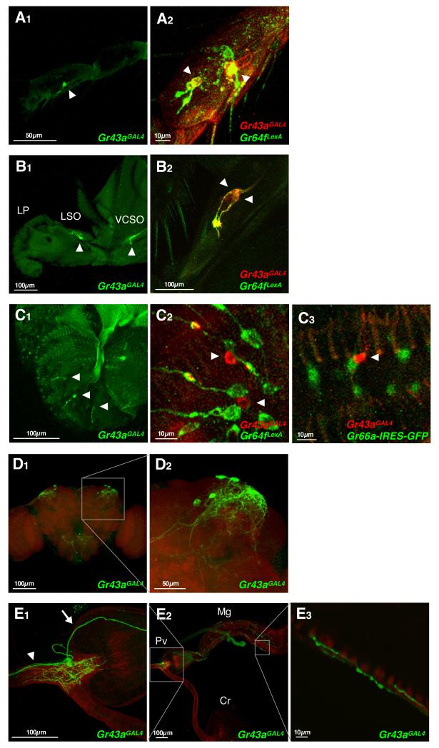
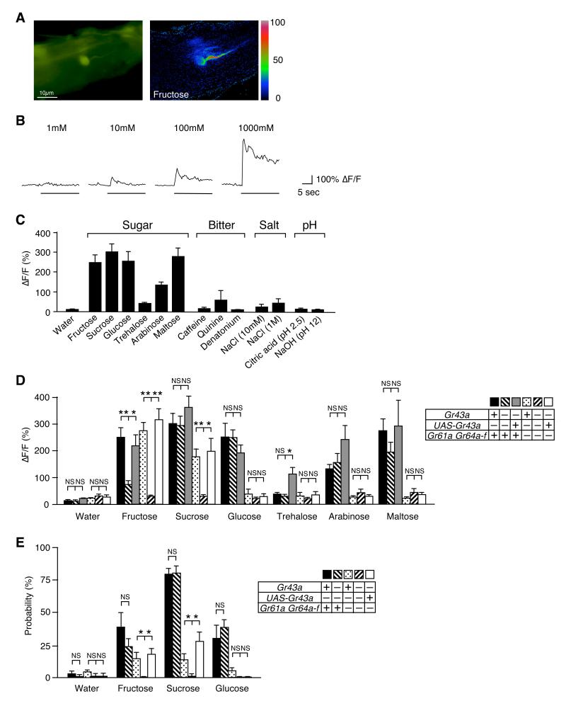
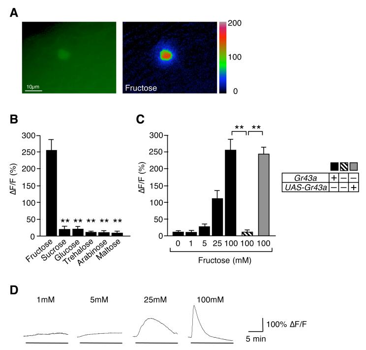
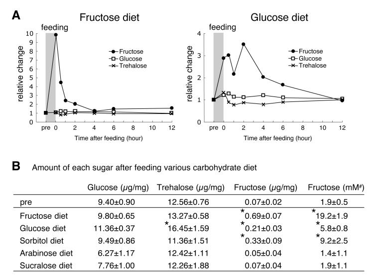
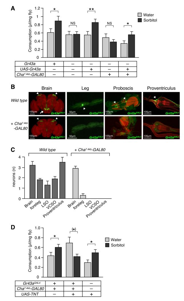
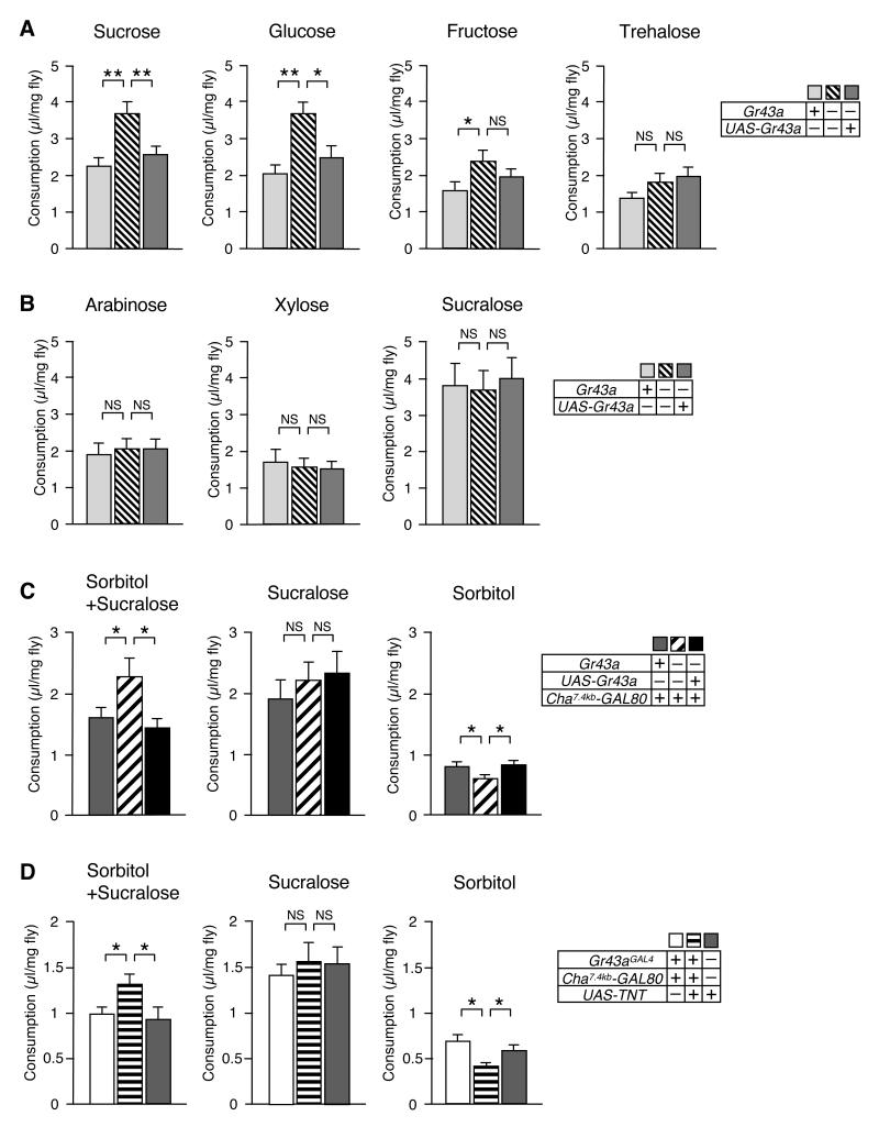
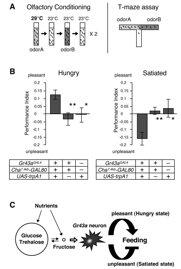
Internal nutrient sensors play important roles in feeding behavior, yet their molecular structure and mechanism of action are poorly understood. Using Ca(2+) imaging and behavioral assays, we show that the gustatory receptor 43a (Gr43a) functions as a narrowly tuned fructose receptor in taste neurons. Remarkably, Gr43a also functions as a fructose receptor in the brain. Interestingly, hemolymph fructose levels are tightly linked to feeding status: after nutritious carbohydrate consumption, fructose levels rise several fold and reach a concentration sufficient to activate Gr43a in the brain. By using different feeding paradigms and artificial activation of Gr43a-expressing brain neurons, we show that Gr43a is both necessary and sufficient to sense hemolymph fructose and promote feeding in hungry flies but suppress feeding in satiated flies. Thus, our studies indicate that the Gr43a-expressing brain neurons function as a nutrient sensor for hemolymph fructose and assign opposing valence to feeding experiences in a satiation-dependent manner.


Figures

Figure 1
Figure 1. Gr43aGAL4 is expressed in chemosensory organs, the brain and the proventriculus
(A-C) Expression of Gr43a in chemosensory organs. Gr43aGAL4 drives strong UAS-mCD8GFP expression in neurons located in the fifth tarsal segment of the foreleg (A1, live GFP), the LSO and the VCSO (B1; live GFP), but only weak expression in the LPs (C1; immunostaining). Co-expression analysis in sweet neurons was performed in flies containing Gr43aGAL4 and Gr64fLexA, driving expression of UAS-mCD8RFP (detected with anti-CD8 antibody) and lexAop-rCD2GFP (detected with anti-GFP antibody), respectively. Co-expression analysis in bitter neurons was performed in flies containing Gr43aGAL4 driving expression of UAS-mCD8RFP (detected with anti-CD8 antibody) and Gr66a-gfp (detected with anti-GFP antibody). LP, labial palp; LSO, labral sensory organ; VCSO, ventral cibarial sense organ. Arrowheads indicate Gr43aGAL4 neurons. (D)Gr43aGAL4 is expressed in 2-4 neurons/hemisphere in the posterior superior lateral protocerebrum. (E)Gr43aGAL4 is expressed in approximately four neurons in the proventricular ganglion. Gr43aGAL4 neurons innervate the lumen of the foregut, but not the crop duct (E2, left inset; E1). Some neurons send projections to the mid gut (E2, right inset; E1, arrow; E3), others to the SOG (E1, arrowhead). Pv, proventriculus; Cr, crop; Mg, mid gut.
Figure 2
Figure 2. GR43a is a fructose receptor
(A) Tarsal neuron expressing G-CaMP3.0 under control of Gr43aGAL4:ΔF pseudocolor fluorescence image was taken 1.5 seconds after application of 100mM fructose (right). (B) G-CaMP3.0 (ΔF/F) fluorescence is dose-dependent (fructose was used as ligand). (C)Gr43aGAL4 neurons responded to sugars, but not to bitter compounds, acid and base. Max ΔF/F within 30 seconds of applications is shown. Sugars and caffeine were at 100mM, quinine and denatonium at 10mM concentration. Error bars represent standard error. 3≤n≤8. (D) Ca2+ response of Gr43aGAL4 neurons with/without Gr43a, Gr61a and Gr64a-f to various sugars. From left to right, genotypes are Gr43aGAL4/+, Gr43aGAL4/Gr43aGAL4, Gr43aGAL4/Gr43aGAL4;UAS-Gr43a, Gr43aGAL4/+;ΔGr61a ΔGr64a-f/ΔGr61a ΔGr64a-f, Gr43aGAL4/Gr43aGAL4Gr61a ΔGr64a-f/ΔGr61a ΔGr64a-f, Gr43aGAL4/Gr43aGAL4;UAS-Gr43aGr61a ΔGr64a-f/ΔGr61a ΔGr64a-f. All sugars are 100mM. NS, not significant; *p < 0.05; **p < 0.0001; ANOVA. Error bars represent standard error. 8≤n≤9. (E)Gr43a is sufficient to induce PER response to fructose. All sugars are 100mM. NS, not significant; *p < 0.05; ANOVA. Error bars represent standard error. 9≤n≤10 experiments (9-21 flies per experiment).
Figure 3
Figure 3. GR43a functions as a fructose sensor in the brain
(A) Brain neuron expressing G-CaMP3.0 under control of Gr43aGAL4: ΔF pseudocolor fluorescence image was taken 24 seconds after application of 100mM fructose (right). (B)Gr43aGAL4 neurons specifically respond to fructose. Max ΔF/F within 15 minutes of application is shown. All sugars are 100 mM. Flies contained two genomic copies of Gr43a. **p < 0.0001; ANOVA. Error bars represent standard error. 6≤n≤7. (C) Response of Gr43aGAL4 neurons to fructose is dose- and Gr43a dependent. **p < 0.0001; ANOVA. Error bars represent standard error. 8≤n≤9. (D) Time-course of G-CaMP3.0 fluorescence changes in Gr43aGAL4 neurons stimulated with different concentrations of fructose.
Figure 4
Figure 4. Metabolic dynamics of circulating sugars
Flies were starved for 24 hours (pre), followed by 40 minutes of feeding. Measurements were performed at indicated times after feeding (see Experimental Procedures). (A) Relative change of internal glucose, trehalose and fructose over time. (B) Amount of glucose, trehalose and fructose (per mg of head tissue) was measured immediately after feeding. *p < 0.05; ANOVA. 5≤n≤12. Last column (#) shows concentration of fructose (converted to mM) using the conservative estimate that 1/5 of the insect mass constitutes hemolymph (Chapman, 1998).
Figure 5
Figure 5. GR43a functions as an internal nutrient sensor
(A) GR43a evaluates nutritional content of carbohydrates. Single flies were subjected to the CAFÉ assay by presenting them with two capillaries containing water and 100mM sorbitol, respectively, for 24 hours. NS, not significant; *p < 0.05; **p < 0.01; ANOVA. Error bars represent standard error. 50≤n≤74. (B)Cha7.4kb-GAL80 restricts Gr43aGAL4 expression to the brain neurons. GFP expression (arrowhead) is missing in all sensory neurons, but remains robust in the brain neurons. Likewise, all projections of sensory neurons to the SOG (arrow) disappeared. (C) Average number of Gr43aGAL4 neurons in different tissues. 3≤n≤11 (without Cha7.4kb-GAL80), 6≤n≤15 (with Cha7.4kb-GAL80). Note that a few legs of flies with Cha7.4kb-GAL80 show very weak GFP expressions. (D)Gr43aGAL4 brain neurons are necessary to evaluate nutritional content of carbohydrates. Flies with silenced Gr43aGAL4 brain neurons (Gr43aGAL4/UAS-TNT; Cha7.4kb-GAL80) lack sorbitol preference, in contrast to control flies. *p < 0.05; ANOVA. Error bars represent standard error. 64≤n≤83.
Figure 6
Figure 6. Gr43a function in brain neurons is sufficient to suppress feeding in satiated flies and promote feeding in hungry flies
Single flies were subjected to the CAFÉ assay for 24 hours by presenting them a single capillary containing the indicated solution. (A)Gr43a suppresses nutritious sugar consumption under satiated conditions. NS, not significant; *p < 0.05; **p < 0.01; ANOVA. Error bars represent standard error. 21≤n≤34. All sugars were used at 100 mM concentration, except sucrose, which was used at 50 mM, to obtain equal nutritional value for mono- and dissacharides. (B) Non-nutritious sugars arabinose (100mM), xylose (100mM) and sucralose (50mM) were consumed in equal amounts by control and Gr43aGAL4 mutant flies. NS, not significant; ANOVA. 24≤n≤27. (C)Gr43aGAL4 brain neurons are sufficient to suppress and promote feeding in satiated and hungry flies, respectively. Cha7.4kb-GAL80 restricts expression of Gr43aGAL4 to the brain (see Figure 5B and 5C). 50mM sucralose was added to “sweeten” sorbitol (100mM), enhance feeding and achieve satiation. For hungry state, sorbitol (100mM) alone was used. NS, not significant; *p < 0.05; ANOVA. 35≤n≤84. (D)Gr43aGAL4 brain neurons are necessary to suppress and promote feeding in satiated and hungry flies, respectively. Flies with silenced Gr43aGAL4 brain neurons (Gr43aGAL4/UAS-TNT; Cha7.4kb-GAL80) consume more nutritious food (sucralose + sorbitol) under satiating conditions, but less under non-satiating conditions (sorbitol alone). NS, not significant; *p < 0.05; ANOVA. 41≤n≤88.
Figure 7
Figure 7. Activation of Gr43aGAL4 brain neurons assigns satiety-dependent valence
(A) Schematic diagram of the olfactory conditioning assay. Flies are exposed to odor A, while their Gr43aGAL4 brain neurons are activated using trpA1 (29°C). After a brief rest period in an odorless vial, they are exposed to odor B in the absence of neural activation (23°C), followed by another rest period in an odorless vial. This training session is repeated once more before the flies are tested in a T-Maize assay for acquired odor preference. (B) Olfactory conditioning assay of starved and satiated flies. Prior conditioning, flies were kept in agarose or agarose containing 250 mM sucrose for 18-24 hours to induce starvation and satiation, respectively. Gr43aGAL4/UAS-trpA1;Cha7.4kb-GAL80/+ flies assign positive valence when hungry (pleasant; left graph), but negative valence when satiated (unpleasant; right graph). Control flies were Gr43aGAL4/+; Cha7.4kb-GAL80/+ and UAS-trpA1/+. *p < 0.05; **p<0.01; ANOVA. 12≤n≤18. (C) Model: The fly’s major blood sugars, glucose and trehalose are kept at a fairly constant, relatively high level. Conversely, internal fructose level is very low, but fluctuates in response to feeding of nutritious sugars. Since nutritious carbohydrates can be converted into fructose, the activity of Gr43aGAL4 brain neurons depends on the nutritious value of the ingested food. Activation of Gr43aGAL4 brain neurons, in combination with the state of satiety, leads either to a pleasant sensation in hungry flies, reinforcing feeding behavior, or is perceived as unpleasant, thereby terminating feeding behavior.

Comment in

  • Let the sensor fit the diet.
    Levin BE. Levin BE. Cell Metab. 2012 Dec 5;16(6):689-90. doi: 10.1016/j.cmet.2012.11.008. Cell Metab. 2012. PMID: 23217252 Free PMC article.

References

    1. Bharucha KN, Tarr P, Zipursky SL. A glucagon-like endocrine pathway in Drosophila modulates both lipid and carbohydrate homeostasis. J Exp Biol. 2008;211:3103–3110. - PMC - PubMed
    1. Buch S, Melcher C, Bauer M, Katzenberger J, Pankratz MJ. Opposing effects of dietary protein and sugar regulate a transcriptional target of Drosophila insulin-like peptide signaling. Cell Metab. 2008;7:321–332. - PubMed
    1. Burke CJ, Waddell S. Remembering nutrient quality of sugar in Drosophila. Curr Biol. 2011;21:746–750. - PMC - PubMed
    1. Cameron P, Hiroi M, Ngai J, Scott K. The molecular basis for water taste in Drosophila. Nature. 2011;465:91–95. - PMC - PubMed
    1. Chapman R. The Insects: Structure and Function. 4th edn Cambridge University Press; Cambridge, New York, Melbourne, Madrid, Cape Town: 1998.

Publication types