Review
. 2012 Nov-Dec;53(6):458-69.

Diastolic filling vortex forces and cardiac adaptations: probing the epigenetic nexus

Affiliations
Review

Diastolic filling vortex forces and cardiac adaptations: probing the epigenetic nexus

Ares Pasipoularides. Hellenic J Cardiol. 2012 Nov-Dec.
No abstract available


Figures

Figure 1
Figure 1
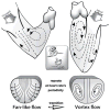
Top: RV/LV diastolic flow patterns: the incoming jet strikes the ventricular apex and sets up a toroidal vortex with its main strength within the outflow tract of each chamber. Center inset: The “vortex-thruster” propulsion mode of cephalopods. Bottom: During the E-wave upstroke, as blood fans away from the central stream toward the endocardial walls, the convective deceleration effect tends, by the Bernoulli mechanism, to raise downstream pressure opposing ventricular inflow; this convective pressure-rise is opposed by the local acceleration gradient, which favors forward flow. During the E-wave downstroke, it is joined in sense and gets reinforced by the now adverse local deceleration gradient and can consequently reverse the flow. This leads to disruption of the boundary between oncoming blood and endocardial walls, or “flow separation,” and to the formation of a toroidal vortex that surrounds a central core.
Figure 2
Figure 2
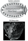
Top: Satisfying the viscous flow “no-slip” condition at a solid-fluid interface, the intraventricular ejection flow-field streamlines are perpendicular to the contracting endocardial surface; consequently, there is no shear (tangential) endocardial stress during ejection. Bottom: Fortuitously, flow-governing fluid dynamic principles do not allow inflow to mirror outflow streamlines throughout the ensuing filling phase—cf. Figure 1, Bottom. During filling, there ensues flow separation and formation of a toroidal vortex that surrounds a central jet. Thus, laminar vortical shear and “squeeze” forces can come into existence.
Figure 3
Figure 3
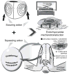
Top: Rotation in the intra-ventricular flow-field can generate a more or less vigorous scouring (tractive shear) of the endocardium lining the chamber. Bottom: Under the action of centripetal and centrifugal forces associated with RV/LV rotatory diastolic flow, the endocardium and other wall components get deformed, akin to a ball squeezed between one’s palms. This dynamic interplay has not been previously recognized in the literature; it and its disturbances are likely to have intriguing epigenetic actions affecting cardiac function and adaptations, acting concurrently with the vortex-induced shear (Top), as summarized in the Middle. Middle: Myocardial cells (endocardium, myocytes, fibroblasts) can move, change shape, and switch genes on and off in response to changes in hydrodynamic shear and “squeeze” (see discussion in text).
Figure 4
Figure 4
During cardiogenesis and in pre- and postnatal life, a circular regulatory pathway exists linking intracardiac flows and associated forces acting on the cardiac walls, to epigenetically influenced mechanosensitive gene expression and changes in the morphology of the developing prenatal or in the phenotype of the adapting postnatal heart. Phenotypic plasticity can, in conjunction with pre- and postnatal operating “environmental” conditions, lead not only to adaptive but also to maladaptive responses and disease. (Modified with permission of PMPH-USA from Pasipoularides A. Heart’s Vortex: Intracardiac Blood Flow Phenomena. Shelton, CT: People’s Medical Publishing House, 2010. 960 pp.)
Figure 5
Figure 5
Summary of epigenetic dynamic actions of RV/LV diastolic toroidal vortex. Cytoskeletal “tensegrity” dynamics create equilibrium between strut compression and string tension, allowing internal structural balance and shape maintenance. Cells sense their physical 3-D “environment,” including variable diastolic vortical shear and “squeeze” forces, by transducing mechanical deformations and forces into differentiated transcription and translation signals, which can adjust cellular and extracellular tissue and organ structure. Mechanosensitive controls modulate myocyte shape and intracellular architecture, and processes as diverse as proliferation, hypertrophy, and apoptosis, involved in cardiac homeostasis and adaptations.

References

    1. Pasipoularides A. Heart’s vortex: intracardiac blood flow phenomena. Shelton, CT: People’s Medical Publishing House; 2010. p. 960. p. See, in particular, Chapter 9. Vortex formation in fluid flow, p. 442–480.
    1. ibid. See, particularly, Chapter 5. Micromanometric, velocimetric, angio- and echocardiographic measurements, p. 234–297, Chapter 10. Cardiac computed tomography, magnetic resonance, and real-time 3-D echocardiography, p. 482–581, and Chapter 11. Postprocessing exploration techniques and display of tomographic data, p. 582–622.

    1. Brutsaert DL. Cardiac endothelial-myocardial signaling: its role in cardiac growth, contractile performance, and rhythmicity. Physiol Rev. 2003;83:59–115. - PubMed
    1. Pasipoularides A, Palacios I, Frist W, Rosenthal S, Newell JB, Powell WJ., Jr Contribution of activation-inactivation dynamics to the impairment of relaxation in hypoxic cat papillary muscle. Am J Physiol. 1985;248(1 Pt 2):R54–62. - PubMed
    1. Pasipoularides A. Fluid dynamic aspects of ejection in hypertrophic cardiomyopathy. Hellenic J Cardiol. 2011;52:416–426. - PMC - PubMed