

Development of a Coxsackievirus A16 neutralization assay based on pseudoviruses for measurement of neutralizing antibody titer in human serum
- PMID: 23178532
- PMCID: PMC7112850
- DOI: 10.1016/j.jviromet.2012.11.014
Development of a Coxsackievirus A16 neutralization assay based on pseudoviruses for measurement of neutralizing antibody titer in human serum
Abstract
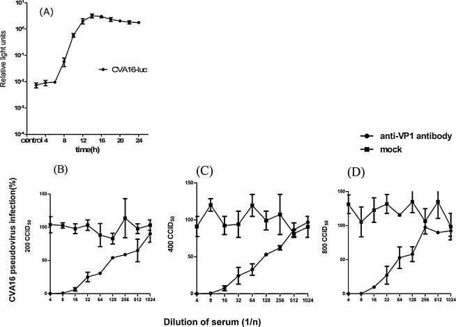
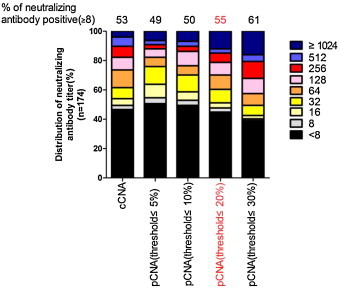
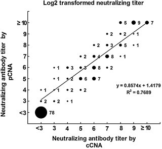
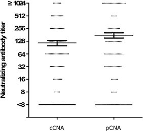
Serum neutralizing antibody titers are indicative of protective immunity against Coxsackievirus A16 (CV-A16) and Enterovirus 71 (EV71), the two main etiological agents of hand, foot and mouth disease (HFMD), and provide the basis for evaluating vaccine efficacy. The current CV-A16 neutralization assay based on inhibition of cytopathic effects requires manual microscopic examination, which is time-consuming and labor-intensive. In this study, a high-throughput neutralization assay was developed by employing CV-A16 pseudoviruses expressing luciferase for detecting infectivity in rhabdomyosarcoma (RD) cells and measuring serum viral neutralizing antibodies. Without the need to use infectious CV-A16 strains, the neutralizing antibody titer against CV-A16 could be determined within 15h by measuring luciferase signals by this assay. The pseudovirus CV-A16 neutralization assay (pCNA) was validated by comparison with a conventional CV-A16 neutralization assay (cCNA) in testing 174 human serum samples collected from children (age <5 years). The neutralizing antibody titers determined by these two assays were well correlated (R(2)=0.7689). These results suggest that the pCNA can serve as a rapid and objective procedure for the measurement of neutralizing antibodies against CV-A16.
Copyright © 2012 Elsevier B.V. All rights reserved.
Figures
Similar articles
-
Development of a high-throughput assay for measuring serum neutralizing antibody against enterovirus 71.J Virol Methods. 2010 Apr;165(1):42-5. doi: 10.1016/j.jviromet.2009.12.015. Epub 2009 Dec 29. J Virol Methods. 2010. PMID: 20036286
-
Development of a poliovirus neutralization test with poliovirus pseudovirus for measurement of neutralizing antibody titer in human serum.Clin Vaccine Immunol. 2011 Nov;18(11):1889-94. doi: 10.1128/CVI.05225-11. Epub 2011 Aug 31. Clin Vaccine Immunol. 2011. PMID: 21880850 Free PMC article.
-
Development of a coxsackievirus A16 neutralization test based on the enzyme-linked immunospot assay.J Virol Methods. 2015 Apr;215-216:56-60. doi: 10.1016/j.jviromet.2015.02.010. Epub 2015 Feb 19. J Virol Methods. 2015. PMID: 25704596
-
Advances in Antigenic Peptide-Based Vaccine and Neutralizing Antibodies against Viruses Causing Hand, Foot, and Mouth Disease.Int J Mol Sci. 2019 Mar 13;20(6):1256. doi: 10.3390/ijms20061256. Int J Mol Sci. 2019. PMID: 30871133 Free PMC article. Review.
-
Prospect and challenges for the development of multivalent vaccines against hand, foot and mouth diseases.Vaccine. 2014 Oct 29;32(47):6177-82. doi: 10.1016/j.vaccine.2014.08.064. Epub 2014 Sep 13. Vaccine. 2014. PMID: 25218294 Review.
Cited by
-
A rapid and quantitative assay for measuring neutralizing antibodies of Coxsackievirus B3.J Virol Methods. 2016 Jun;232:1-7. doi: 10.1016/j.jviromet.2016.02.010. Epub 2016 Mar 4. J Virol Methods. 2016. PMID: 26947399 Free PMC article.
-
Coxsackievirus A6 2C protein antagonizes IFN-β production through MDA5 and RIG-I depletion.J Virol. 2023 Nov 30;97(11):e0107523. doi: 10.1128/jvi.01075-23. Epub 2023 Oct 17. J Virol. 2023. PMID: 37847581 Free PMC article.
-
Identification of luteolin as enterovirus 71 and coxsackievirus A16 inhibitors through reporter viruses and cell viability-based screening.Viruses. 2014 Jul 17;6(7):2778-95. doi: 10.3390/v6072778. Viruses. 2014. PMID: 25036464 Free PMC article.
-
Coxsackievirus A16: epidemiology, diagnosis, and vaccine.Hum Vaccin Immunother. 2014;10(2):360-7. doi: 10.4161/hv.27087. Epub 2013 Nov 14. Hum Vaccin Immunother. 2014. PMID: 24231751 Free PMC article. Review.
-
Pseudotyped Viruses for Enterovirus.Adv Exp Med Biol. 2023;1407:209-228. doi: 10.1007/978-981-99-0113-5_11. Adv Exp Med Biol. 2023. PMID: 36920699
References
-
- Ang L.W., Koh B.K., Chan K.P., Chua L.T., James L., Goh K.T. Epidemiology and control of hand, foot and mouth disease in Singapore, 2001–2007. Ann. Acad. Med. Singapore. 2009;38:106–112. - PubMed
-
- Bartosch B., Bukh J., Meunier J.C., Granier C., Engle R.E., Blackwelder W.C., Emerson S.U., Cosset F.L., Purcell R.H. In vitro assay for neutralizing antibody to hepatitis C virus: evidence for broadly conserved neutralization epitopes. Proc. Natl. Acad. Sci. U.S.A. 2003;100:14199–14204. - PMC - PubMed
-
- Chang L.Y., King C.C., Hsu K.H., Ning H.C., Tsao K.C., Li C.C., Huang Y.C., Shih S.R., Chiou S.T., Chen P.Y., Chang H.J., Lin T.Y. Risk factors of enterovirus 71 infection and associated hand, foot, and mouth disease/herpangina in children during an epidemic in Taiwan. Pediatrics. 2002;109:e88. - PubMed
-
- Chang L.Y., Lin T.Y., Huang Y.C., Tsao K.C., Shih S.R., Kuo M.L., Ning H.C., Chung P.W., Kang C.M. Comparison of enterovirus 71 and coxsackie-virus A16 clinical illnesses during the Taiwan enterovirus epidemic, 1998. Pediatr. Infect. Dis. J. 1999;18:1092–1096. - PubMed
Publication types
MeSH terms
Substances
LinkOut - more resources
Full Text Sources
Other Literature Sources
Medical
Miscellaneous
