. 2013 Jan 9;32(1):112-25.
doi: 10.1038/emboj.2012.304. Epub 2012 Nov 23.

APLF promotes the assembly and activity of non-homologous end joining protein complexes

Affiliations

APLF promotes the assembly and activity of non-homologous end joining protein complexes

Gabrielle J Grundy et al. EMBO J. .

Abstract

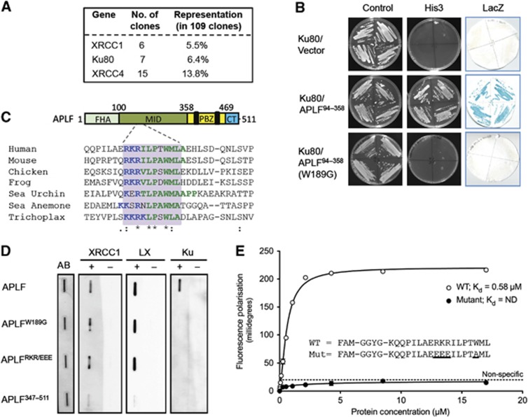
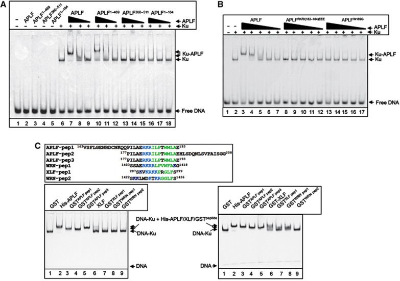
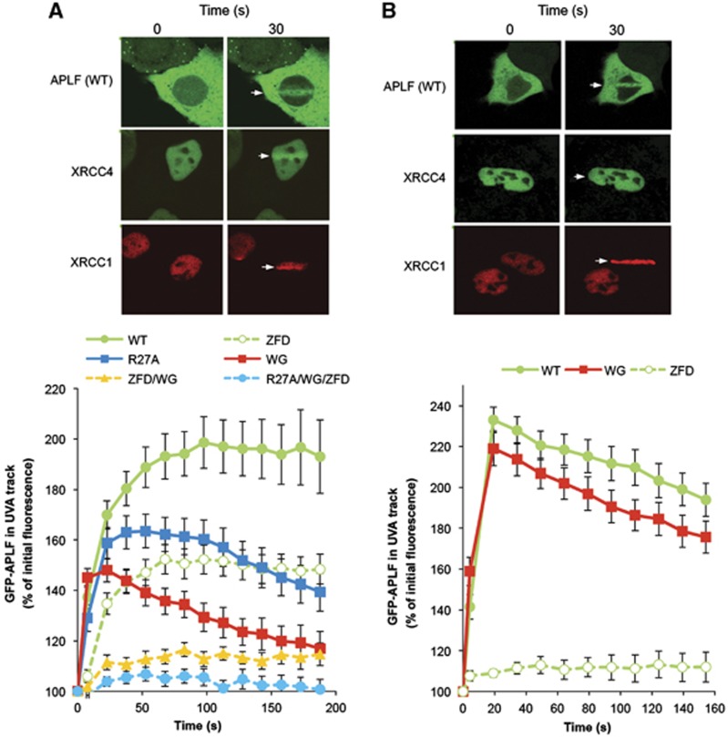
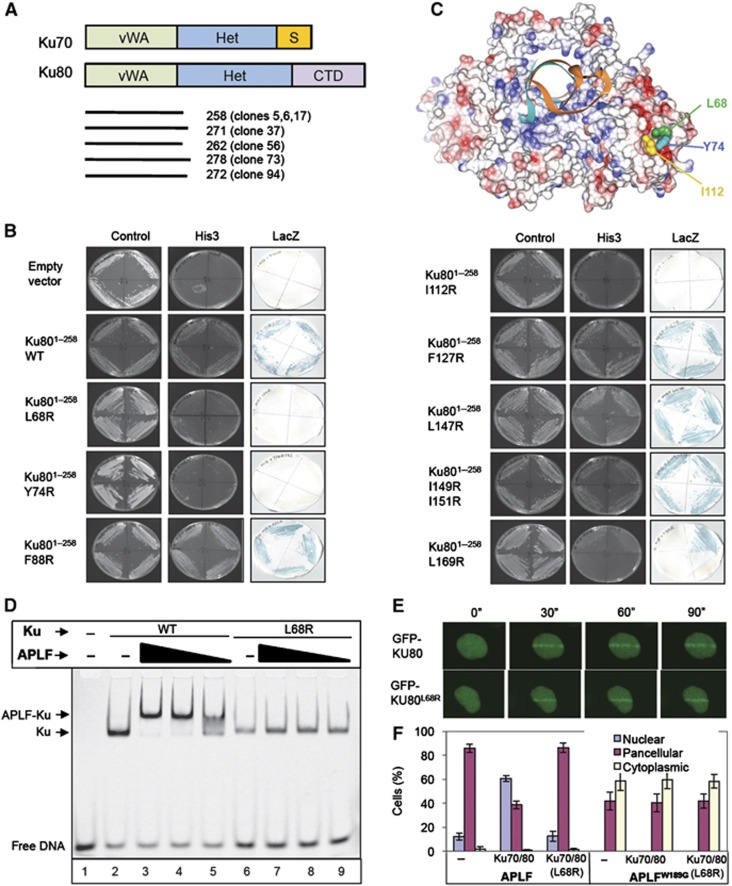
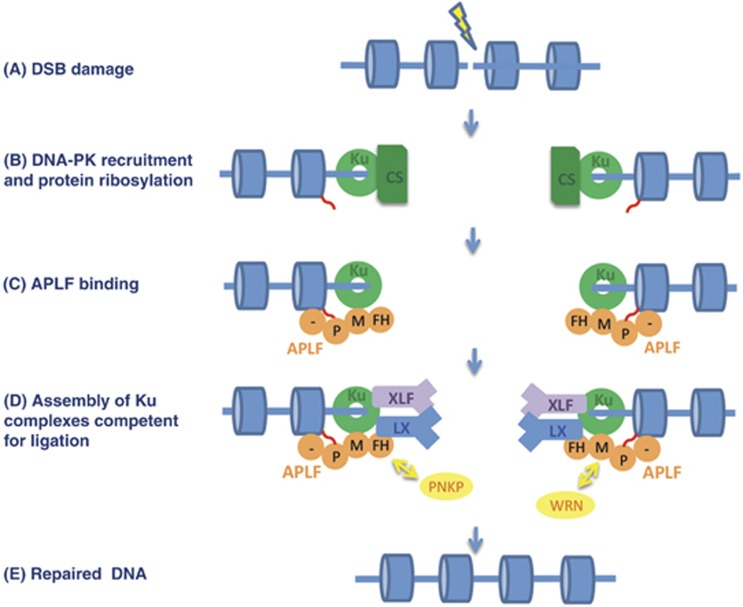
Non-homologous end joining (NHEJ) is critical for the maintenance of genetic integrity and DNA double-strand break (DSB) repair. NHEJ is regulated by a series of interactions between core components of the pathway, including Ku heterodimer, XLF/Cernunnos, and XRCC4/DNA Ligase 4 (Lig4). However, the mechanisms by which these proteins assemble into functional protein-DNA complexes are not fully understood. Here, we show that the von Willebrand (vWA) domain of Ku80 fulfills a critical role in this process by recruiting Aprataxin-and-PNK-Like Factor (APLF) into Ku-DNA complexes. APLF, in turn, functions as a scaffold protein and promotes the recruitment and/or retention of XRCC4-Lig4 and XLF, thereby assembling multi-protein Ku complexes capable of efficient DNA ligation in vitro and in cells. Disruption of the interactions between APLF and either Ku80 or XRCC4-Lig4 disrupts the assembly and activity of Ku complexes, and confers cellular hypersensitivity and reduced rates of chromosomal DSB repair in avian and human cells, respectively. Collectively, these data identify a role for the vWA domain of Ku80 and a molecular mechanism by which DNA ligase proficient complexes are assembled during NHEJ in mammalian cells, and reveal APLF to be a structural component of this critical DSB repair pathway.


Conflict of interest statement

The authors declare that they have no conflict of interest.

Figures

Figure 1
Figure 1
Identification of a novel peptide motif in APLF that binds Ku80. (A) Recovery of human cDNA clones encoding XRCC1, XRCC4, or Ku80 in a yeast 2-hybrid (Y2H) screen employing APLF as bait. The number and relative representation of each clone type is indicated. (B) Ku80 interacts with APLF residues 94–358, and requires W189 for this interaction. Y190 budding yeast harbouring pACT-Ku80 (library clone 5) and empty pGBKT7, pGBKT7-APLF94–358, or pGBKT7-APLFW189G was examined for activation of the His3 and LacZ reporter genes by Y2H analysis. (C) Schematic of APLF depicting a conserved Ku80-binding peptide motif in the MID domain. Top, APLF cartoon depicting the interaction domains; FHA domain that interacts with XRCC1 and XRCC4 (FHA), MID domain that interacts with Ku80, PBZ domain that interacts with poly (ADP-ribose), and C-terminal (CT) domain that interacts with histone H3/H4. Bottom, ClustalW alignment of the putative Ku80-binding peptide motif (grey box) from the indicated organisms. The NCBI accession numbers employed are NP_775816, NP_001163960, XP_419335, NM_001113131, XP_788055, XP_001635068, and XP_002117261. (D) Mutation of the conserved peptide motif in APLF prevents Ku binding, in vitro. One microgram of wild-type APLF, APLFW189G, APLFRKR(182–184)EEE, or APLF347–511 was slot blotted onto nitrocellulose and individual membrane strips stained with Amido black as a loading control (AB) or mock incubated (−) or incubated (+) with 100 nM of recombinant XRCC1, XRCC4-Lig4 heterodimer (LX), or Ku70/80 as indicated. (E) The APLF conserved peptide motif is sufficient to bind Ku. The indicated fluorescein-labelled peptides spanning the wild-type (WT) or mutant APLF conserved peptide motif were examined for binding to Ku70/80ΔC by fluorescence polarisation. Data points are the average of three independent experiments, with error bars representing one standard deviation.
Figure 2
Figure 2
The Ku-binding motif is a novel autonomous module for recruiting proteins into Ku-DNA complexes. (A) The MID domain is required for APLF recruitment into DNA complexes containing Ku. A Cy3-labelled 30-bp duplex oligonucleotide (10 nM) harbouring blunt-ended termini was incubated with (+) or without (−) recombinant Ku70/80 (20 nM) in the absence (−) or presence of 150, 50, or 17 nM of the indicated recombinant APLF protein and employed in EMSA. Where a single concentration of APLF is indicated, the protein was employed at 150 nM. The composition of the protein DNA complexes is indicated on the right (arrows). (B) The Ku-binding motif is required for APLF recruitment into DNA complexes containing Ku. EMSAs were conducted as above. Where indicated, Ku70/80 was present at 20 nM and the indicated wild-type and mutant APLF proteins were employed at 150, 50, 17, 6 and 2 nM. (C) The Ku-binding motif is an autonomous module for protein recruitment into Ku-DNA complexes. Top left, alignment of the peptide sequences from APLF, XLF, and WRN that were fused to GST and analysed in the bottom panels, by EMSA. The conserved basic and hydrophobic patches that characterise the Ku-binding motif are indicated in blue and green, respectively. Bottom left, Cy3-labelled 30-bp duplex oligonucleotide (10 nM) was incubated with Ku70/80 (10 nM) in the presence of 0.5 μM of GST, the indicated GST peptides, full-length His-APLF (lane 2), or full-length XLF (lane 6), and employed in EMSAs as described above. Bottom right, Cy3-labelled 30-bp duplex oligonucleotide was incubated as above in the presence of GST (3 μM), His-APLF (0.5 μM), GST-XLF (1.6 μM), or the indicated GST-APLF (1.3 μM), GST-XLF (1.6 μM), or GST-WRN (3 μM) peptides.
Figure 3
Figure 3
The Ku80-binding motif promotes APLF accumulation at chromosome damage. A549 cells were transiently transfected with constructs encoding wild-type YFP-APLF (WT) or YFP-APLF harbouring point mutations in the PBZ domain (YFP-APLFZFD; ‘ZFD’), FHA domain (YFP-APLFR27A; ‘R27A’), Ku-binding motif (YFP-APLFW189G; ‘WG’), or in combinations of these (‘ZFD/WG’, ‘R27A/WG/ZFD’). mRFP-XRCC1 and GFP-XRCC4 were used as markers of recruitment to single- and double-strand breaks, respectively. Cells were irradiated with 4.36 J/m2 (A) or 0.22 J/m2 (B) using a UVA laser (arrow). Images were captured at 15 s intervals after laser irradiation. For each data point, data are normalised to the YFP fluorescence intensity prior to irradiation (set to 100%). Data are the mean (±s.e.m.) of 10 or more individual cells per data point. Representative examples of YFP-APLF, GFP-XRCC4, and mRFP-XRCC1 accumulation at sites of UVA laser damage are shown (top).
Figure 4
Figure 4
APLF interacts with the vWA domain of Ku80. (A) Cartoon of Ku70 and Ku80, depicting the von Willebrand-like (‘vWA’) domains, heterodimerisation domains (‘Het’), Ku70 SAP domain (‘S’), and Ku80 C-terminal domain (‘CTD’). The regions of Ku80 recovered by APLF in the Y2H screen depicted in Figure 1A are shown (bottom). (B) Mutation of the Ku80 vWA domain disrupts interaction with APLF. Y190 cells harbouring empty pGBKT7 or pGBKT7-APLF94–358 (encoding the MID domain) and either empty pACT2 or the indicated wild-type or mutant derivative of pACT-Ku801–258 (clone 5) were examined for His3 and LacZ reporter gene expression. (C) Residues required for APLF interaction co-localise in a hydrophobic interface on the surface of the Ku80 vWA domain. The location of L68, Y74, and I112 within the Ku heterodimer (RCSB PDB entry; 1JEY) (Walker et al, 2001) is shown. Blue and red denote basic and acidic regions, respectively. (D) Mutation of the Ku80 vWA domain prevents recruitment of APLF into DNA complexes containing Ku. A Cy3-labelled 30-bp duplex (10 nM) was incubated with (+) or without (−) 10 nM wild-type Ku (Ku70/80Δ) or mutant Ku (Ku70/80ΔL68R) in the absence (−) or presence of 700, 350, or 175 nM of the indicated recombinant APLF protein and employed in EMSA. (E) Normal accumulation of Ku70/GFP-Ku80L68R at sites of chromosome damage. A549 cells were transiently co-transfected with His-Ku70 and either GFP-Ku80 or GFP-Ku80L68R prior to UVA laser irradiation. Images were captured at 30 s intervals after laser irradiation. (F) Impact of mutations in the vWA domain on the subcellular localisation of APLF. mRFP localisation following co-transfection with mRFP-APLF or mRFP-APLFW189G and GFP vector, GFP-Ku70/GFP-Ku80, or GFP-Ku70/GFP-Ku80L68R. Data are the mean of three independent experiments (±s.e.m.).
Figure 5
Figure 5
APLF promotes NHEJ complex assembly. (A) Co-assembly of APLF and XRCC4-Lig4 into 19-bp DNA complexes containing Ku. A Cy3-labelled 19-bp duplex (10 nM) was incubated with Ku70/80ΔC (20 nM), in the absence or presence of APLF (0.4 μM) and/or XRCC4-Lig4 (LX; 0.4 μM) and then employed in EMSA. (B) NHEJ protein complexes assembled by APLF are super-shifted by anti-APLF and anti-XRCC4 antibodies. A Cy3-labelled 19-bp duplex was incubated with Ku70/80ΔC as described above in the presence or absence of APLF (0.4 μM) and/or XRCC4-Lig4 (LX; 0.55 μM) and/or the indicated anti-XRCC4 (Santa Cruz; sc-8285) or anti-APLF (Abmart 2G11) antibody and employed in EMSA. (C) Co-assembly of APLF and XRCC4-Lig4 into 60-bp DNA complexes containing Ku. A Cy3-labelled 60-bp duplex (10 nM) was incubated with full-length Ku70/80 (20 nM) in the presence and absence of APLF (0.4 μM) and 0–140 nM XRCC4-Lig4 (LX), as indicated. (D) APLF promotes assembly of both XRCC4-Lig4 and XLF into Ku-DNA complexes and functions as a molecular scaffold. A Cy3-labelled 19-bp duplex (10 nM) was incubated in the absence or presence of full-length Ku70/80 (20 nM), XRCC4-Lig4 (LX; 0.2 μM), XLF (1 μM), and the indicated wild-type or mutant APLF (0.2 μM), and then employed in EMSA.
Figure 6
Figure 6
APLF promotes the recruitment and/or retention of XRCC4-Lig4 and XLF at sites of chromosome damage. (A) APLF is not required for recruitment/retention of Ku at sites of UVA damage. A549 cells mock depleted (WT) or stably depleted of APLF (APLF KD) were transiently transfected with expression constructs encoding GFP-Ku70 and GFP-Ku80 and images were captured at the indicated times after laser UVA micro-irradiation. For each data point, data are normalised to the GFP fluorescence intensity prior to irradiation (set to 100%). Data are the mean (±s.e.m.) of 10 or more individual cells. (B) APLF promotes recruitment/retention of GFP-XRCC4 at sites of UVA damage. APLF KD cells were transiently transfected with GFP-XRCC4 and either mRFP vector (V) or shRNA-resistant mRFP-APLF (WT), mRFP-APLFW189G (WG), or mRFP-APLFR27A (R27A), and analysed as described above. (C) APLF promotes recruitment/retention of GFP-XLF at sites of UVA damage. APLF KD cells were co-transfected with GFP-XLF and mRFP vector (V), mRFP-APLF (WT), mRFP-APLFW189G (WG), or mRFP-APLFR27A (R27A), and analysed as described above.
Figure 7
Figure 7
APLF scaffold activity stimulates DNA ligation in vitro and is required for rapid repair of, and resistance to, chromosomal DSBs. (A) APLF stimulates XRCC4-Lig4 activity in vitro. A Cy3-labelled 247-bp DNA duplex with one ligatable end was incubated for 30 min with recombinant XRCC4-Lig4 (10 nM) alone (‘buffer’) or in the presence or absence as indicated of recombinant Ku70/80 (‘Ku’; 20 nM), wild-type APLF (‘WT’), APLFW189G (‘WG’), APLFR27A (‘RA’), or APLFW189G/R27A (‘RA/WG’). Reaction products were fractionated by denaturing PAGE. A representative image from one experiment is presented (top), along with quantitative data from four independent experiments showing the mean (±s.e.m.) fraction of total DNA converted to a 494-bp product (bottom). (B) APLF scaffold activity is required to accelerate DSB repair. A549 cells were transiently co-transfected with empty pSUPER (‘Mock’) or pSUPER-APLF (‘APLF KD’) and either empty pcD2E expression vector (‘V’) or pcD2E encoding shRNA-resistant wild-type APLF (‘WT’), APLFR27A (‘R27A’), or APLFW189G (‘W189G’). Transfected cells were mock irradiated or γ irradiated (2 Gy) and allowed to recover for the times indicated prior to immunostaining. Data are the mean number (±s.e.m.) of γH2AX foci scored in G1 cells from three independent experiments. Inset, immunoblotting for levels of APLF and histone H1 (loading control) in untreated cells and γ-irradiated cells (30 min after irradiation). (C) Reduced plasmid re-joining in APLF−/− DT40 cells. Wild-type (Clone 18) and APLF−/− DT40 cells were co-transfected with circular RFP and linear GFP vector, 18 h before analysis by FACS. Data are the percentage of RFP-positive cells expressing GFP and are the mean (±s.e.m.) of five independent experiments. **P<0.01 by Student’s t-test. (D) Hypersensitivity of APLF−/− DT40 cells to γ radiation. Wild-type DT40 cells (clone 18), APLF−/− DT40 cells, and APLF−/− DT40 cells stably transfected with empty expression vector or with expression construct encoding myc-tagged wild-type (WT) human APLF or the indicated myc-tagged mutant human APLF were γ irradiated with the indicated dose and cell colonies counted 10 days after treatment. Inset, anti-myc Western blot depicting expression levels of the indicated recombinant APLF protein. Note that the APLF proteins expressed in these experiments were additionally tagged with an SV40 nuclear localisation signal.
Figure 8
Figure 8
A model for APLF scaffold activity during NHEJ. (A) DSB induction. (B) Ku heterodimer binds the DSB and recruits DNA-PKcs, and PARP3 ribosylates (red wavy lines) nucleosomes (blue cylinders) near to the break. (C) APLF (orange) binds ribosylated nucleosomes via its C-terminal poly (ADP-ribose)-binding domain (PBZ domain; ‘P’) and histone-binding acidic tail, and binds Ku heterodimer via its Ku80-binding MID domain (‘M’). (D) Binding and retention of the DNA ligase co-factors XLF and XRCC4-Lig4 is promoted by their interaction with both Ku and APLF, forming a ‘holocomplex’ competent for ligation. We suggest that this complex is highly dynamic, allowing exchange between different NHEJ proteins and individual APLF domains as-and-when required. For example, APLF might be replaced by PNKP during DNA end processing, should the latter detect the presence of its DNA substrate, via interchange between their respective XRCC4-binding FHA domains. Similarly, APLF may exchange with WRN, via interchange between their respective Ku80-binding motifs. (E) Following end processing (and gap filling if required), XLF and XRCC4-Lig4 ligate the DSB.

References

    1. Ahel I, Ahel D, Matsusaka T, Clark AJ, Pines J, Boulton SJ, West SC (2008) Poly(ADP-ribose)-binding zinc finger motifs in DNA repair/checkpoint proteins. Nature 451: 81–85 - PubMed
    1. Ahnesorg P, Smith P, Jackson SP (2006) XLF interacts with the XRCC4-DNA ligase IV complex to promote DNA nonhomologous end-joining. Cell 124: 301–313 - PubMed
    1. Aravind L, Koonin EV (2001) Prokaryotic homologs of the eukaryotic DNA-end-binding protein Ku, novel domains in the Ku protein and prediction of a prokaryotic double-strand break repair system. Genome Res 11: 1365–1374 - PMC - PubMed
    1. Arosio D, Cui S, Ortega C, Chovanec M, Di Marco S, Baldini G, Falaschi A, Vindigni A (2002) Studies on the mode of Ku interaction with DNA. J Biol Chem 277: 9741–9748 - PubMed
    1. Bekker-Jensen S, Fugger K, Danielsen JR, Gromova I, Sehested M, Celis J, Bartek J, Lukas J, Mailand N (2007) Human Xip1 (C2orf13) is a novel regulator of cellular responses to DNA strand breaks. J Biol Chem 282: 19638–19643 - PubMed

Publication types

MeSH terms