

Restriction of intestinal stem cell expansion and the regenerative response by YAP
- PMID: 23178811
- PMCID: PMC3536889
- DOI: 10.1038/nature11693
Restriction of intestinal stem cell expansion and the regenerative response by YAP
Abstract
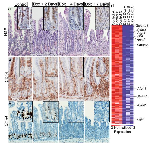
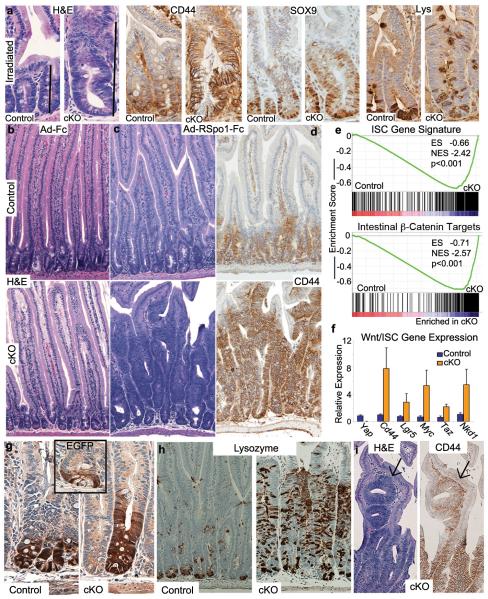
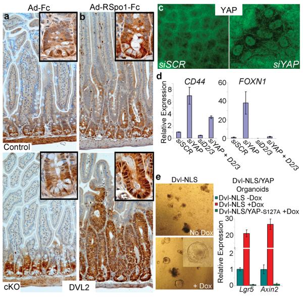
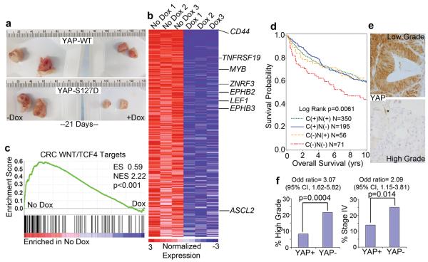
A remarkable feature of regenerative processes is their ability to halt proliferation once an organ's structure has been restored. The Wnt signalling pathway is the major driving force for homeostatic self-renewal and regeneration in the mammalian intestine. However, the mechanisms that counterbalance Wnt-driven proliferation are poorly understood. Here we demonstrate in mice and humans that yes-associated protein 1 (YAP; also known as YAP1)--a protein known for its powerful growth-inducing and oncogenic properties--has an unexpected growth-suppressive function, restricting Wnt signals during intestinal regeneration. Transgenic expression of YAP reduces Wnt target gene expression and results in the rapid loss of intestinal crypts. In addition, loss of YAP results in Wnt hypersensitivity during regeneration, leading to hyperplasia, expansion of intestinal stem cells and niche cells, and formation of ectopic crypts and microadenomas. We find that cytoplasmic YAP restricts elevated Wnt signalling independently of the AXIN-APC-GSK-3β complex partly by limiting the activity of dishevelled (DVL). DVL signals in the nucleus of intestinal stem cells, and its forced expression leads to enhanced Wnt signalling in crypts. YAP dampens Wnt signals by restricting DVL nuclear translocation during regenerative growth. Finally, we provide evidence that YAP is silenced in a subset of highly aggressive and undifferentiated human colorectal carcinomas, and that its expression can restrict the growth of colorectal carcinoma xenografts. Collectively, our work describes a novel mechanistic paradigm for how proliferative signals are counterbalanced in regenerating tissues. Additionally, our findings have important implications for the targeting of YAP in human malignancies.
Figures
Comment in
-
Development: resizing the guts.Nat Rev Mol Cell Biol. 2013 Jan;14(1):4. doi: 10.1038/nrm3501. Epub 2012 Dec 12. Nat Rev Mol Cell Biol. 2013. PMID: 23232564 No abstract available.
-
Keeping stem cells in check: a hippo balancing act.Cell Stem Cell. 2013 Jan 3;12(1):3-5. doi: 10.1016/j.stem.2012.12.004. Cell Stem Cell. 2013. PMID: 23290131
References
-
- Camargo FD, et al. YAP1 increases organ size and expands undifferentiated progenitor cells. Curr Biol. 2007;17:2054–60. - PubMed
Publication types
MeSH terms
Substances
Associated data
- Actions
Grants and funding
- P01CA87969/CA/NCI NIH HHS/United States
- AR064036/AR/NIAMS NIH HHS/United States
- R01 CA151993/CA/NCI NIH HHS/United States
- U24 DK085532/DK/NIDDK NIH HHS/United States
- R01 CA131426/CA/NCI NIH HHS/United States
- U01 DK085527/DK/NIDDK NIH HHS/United States
- P50CA127003/CA/NCI NIH HHS/United States
- R01 DK091427/DK/NIDDK NIH HHS/United States
- U01 DK085532/DK/NIDDK NIH HHS/United States
- 1U01DK085527/DK/NIDDK NIH HHS/United States
- P50 CA127003/CA/NCI NIH HHS/United States
- R01 AR064036/AR/NIAMS NIH HHS/United States
- 1K08DK096048/DK/NIDDK NIH HHS/United States
- K08 DK096048/DK/NIDDK NIH HHS/United States
- P30 DK049216/DK/NIDDK NIH HHS/United States
LinkOut - more resources
Full Text Sources
Other Literature Sources
Medical
Molecular Biology Databases
Research Materials
