Review
. 2013 Sep;70(17):3089-108.
doi: 10.1007/s00018-012-1212-1. Epub 2012 Nov 22.

AID and Apobec3G haphazard deamination and mutational diversity

Affiliations
Review

AID and Apobec3G haphazard deamination and mutational diversity

Malgorzata Jaszczur et al. Cell Mol Life Sci. 2013 Sep.

Abstract

Activation-induced deoxycytidine deaminase (AID) and Apobec 3G (Apo3G) cause mutational diversity by initiating mutations on regions of single-stranded (ss) DNA. Expressed in B cells, AID deaminates C → U in actively transcribed immunoglobulin (Ig) variable and switch regions to initiate the somatic hypermutation (SHM) and class switch recombination (CSR) that are essential for antibody diversity. Apo3G expressed in T cells catalyzes C deaminations on reverse transcribed cDNA causing HIV-1 retroviral inactivation. When operating properly, AID- and Apo3G-initiated mutations boost human fitness. Yet, both enzymes are potentially powerful somatic cell "mutators". Loss of regulated expression and proper genome targeting can cause human cancer. Here, we review well-established biological roles of AID and Apo3G. We provide a synopsis of AID partnering proteins during SHM and CSR, and describe how an Apo2 crystal structure provides "surrogate" insight for AID and Apo3G biochemical behavior. However, large gaps remain in our understanding of how dC deaminases search ssDNA to identify trinucleotide motifs to deaminate. We discuss two recent methods to analyze ssDNA scanning and deamination. Apo3G scanning and deamination is visualized in real-time using single-molecule FRET, and AID deamination efficiencies are determined with a random walk analysis. AID and Apo3G encounter many candidate deamination sites while scanning ssDNA. Generating mutational diversity is a principal aim of AID and an important ancillary property of Apo3G. Success seems likely to involve hit and miss deamination motif targeting, biased strongly toward miss.


Figures

Fig. 1
Fig. 1
AID-targeted deamination at V(D)J and S regions of Ig genes initiates SHM and CSR. Low affinity IgM-producing IgVH genes undergo SHM and CSR which require C → U deamination by AID at V(D)J and switch (S) regions, respectively. Transcription from the V promoter (P) is needed for AID access to V(D)J regions, whereas germline transcription from the I promoters is required for AID targeting to the donor switch () and a downstream acceptor S (shown in this case). Error-prone processing of G:U mispair by MMR, BER and replication introduces a huge number of mutations (~10−4 to 10−3 per base per generation) in the antigen-binding V(D)J region as well as the rejoining Sμ–Sε regions. CSR combines the V(D)J exon with one of the appropriate downstream constant C regions γ3, (γ1, γ2b, and γ2a not shown), ε or α, converting IgM or IgD to the other isotypes IgG3 (IgG1, IgG2b, and IgG2a not shown), IgE, or IgA. The graph on top shows a typical SHM profile. Mutations start ~150 bp downstream from the promoter (P) at the leader sequence (L) reaching a maximum frequency over the V(D)J coding exon decaying exponentially towards the 3′ end. is an intronic enhancer. E3′s are enhancers at 3′ regulatory regions (3′RR)
Fig. 2
Fig. 2
Apo3G-dependent restriction of HIV-1. Apo3G is encapsidated into Vif-deficient (∆vif) HIV-1 virions. Upon infection, Apo3G enters the cytoplasm of the targeted T cell and deaminates multiple C on the HIV-1 reverse transcribed minus (−) cDNA strand creating a massive number of U residues. Synthesis of the (+) strand, using U as templates, introduces hypermutation in essential genes, effectively inactivating HIV-1 infectivity. Alternatively, abasic sites resulted from the removal of U by the cellular enzyme, uracil DNA glycosylase, could inhibit the synthesis of the HIV-1 (+) strand cDNA or could serve as substrates for apurinic-apyrimidinic endonucleases leading to degradation of the cDNA reverse transcribed intermediate
Fig. 3
Fig. 3
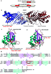
Apobec protein domains, structures and sequence alignments. a AID and six other Apobec’s contain a single Zn2+ coordinating deoxycytidine/cytidine deaminase domain, whereas four other members are double-domain deaminases. The N-terminal domain (CD1) of Apo3G, Apo3F, and Apo3DE is inactive while the C-terminal domain (CD2) is catalytically active. b The crystal structure of Apo2 tetramer (Protein Data Bank 2NYT). Head-to-head interaction of two Apo2 dimers is mediated by tetramer interface amino acid residues of α-helix 6, active center loop1, and flexible loop 7. The Apo2 dimer is formed by combining two β2 strands to make one wide β-sheet structure (indicated by arrow). Each monomer is highlighted in a different color. The monomer active site residues C128, C131, and H98 (red) coordinate Zn2+ molecule (red). c The crystal structure of the Apo3G-CD2 (Protein Data Bank 3E1U). The α-helixes are marked in blue and β-strands in pink. The C288, C291, and H257 (red) coordinate Zn2+ molecule (red), whereas E259 (red) acts as a proton shuffler during the hydrolytic deamination reaction. d The AID model was generated using Apo3G-CD2 as a template. Active site residues H56, E58, C87, and C90 (red) are important for catalysis. H56, C87, and C90 are involved in coordinating the Zn ion (red sphere) while E58 is involved in proton shuffling. Predicted residues at the dimeric and tetrameric interfaces are indicated by arrows. e Alignment of AID, Apo3G-CD2, and Apo2. HIGM-2 base substitution mutations and C-terminal deletion mutations are indicated on the top. The amino acids from the protein active site necessary for catalysis are marked in red. The specificity loop is marked in red both in the Apo3G-CD2 structure and amino acid alignment
Fig. 4
Fig. 4
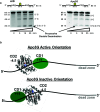
Model for Apo3G deamination polarity. a A Fluorescein (F)-labeled 85-nt ssDNA containing 2 identical deamination motifs (AGC for AID, left and CCC for Apo3G, right) was incubated with AID or Apo3G. Following treatment with Uracil-DNA Glycosylase and hot alkali, single deaminations at the 5′ site (5′C) or the 3′ site (3′C) are detected as the appearance of the labeled 67-nt or 48-nt fragment, respectively. Processive deamination at both the 5′ and 3′ sites (5′C and 3′C) on the same substrate is detected as the presence of a 30-nt fragment. AID deaminates 5′ and 3′ target motifs with equal efficiency (left gel), whereas Apo3G deaminatates the 5′ target motif preferentially (right gel). b Proposed model for Apo3G deamination polarity. Apo3G can bind ssDNA, with equal probability, in active or inactive orientations. In the active orientation, the active domain CD2 (charge = −4.5) is positioned toward the 5′-end and CD1 (+11) is facing the 3′-end. In the inactive orientation, Apo3G residues responsible for substrate specificity are distant from the 5′-CCC target motif and away from zinc ion, thereby precluding catalysis. The inability of the CD2 domain to bind to 30 nt at the 3′end in the active orientation results in a “deamination dead zone”
Fig. 5
Fig. 5
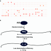
Stochastic AID-catalyzed deamination. a Diverse AID deamination patterns on individual substrates containing 30 AAC (red dot) and AGC (orange dot) hot deamination motifs. AID deamination at the target motifs is shown as T [17]. The representative clones contain 2–10 mutations, distributed as singletons or clusters (doublets, triplets, etc.). b Random walk model depicting AID scanning and catalyzing inefficient deamination of C to U on ssDNA [17]. AID binds to ssDNA randomly and slides in both direction, while catalyzing C deaminations processively. The sliding/hopping distance is determined by a geometric distribution. A good fit of the model to the data occurs when AID slides/hops for average distance of 10 motifs (30 nt), but only rarely deaminates C to U, at about 3 % efficiency for even “hottest” AAC motifs, thus ensuring mutational diversity [17]
Fig. 6
Fig. 6
Single-molecule microscopy to visualize Apo3G scanning on ssDNA. a Total internal reflectance fluorescence (TIRF) microscope set up for analyzing Apo3G motion along ssDNA. ssDNA (92 nt) is annealed to a Cy3-labeled (D donor), surface immobilized anchor DNA. Binding and scanning of Cy5-labeled (A acceptor) Apo3G results in FRET changes. b, c The representative smFRET Scanning Trajectories for poly dT substrate with a 5′ hot CCC motif (pdT 5′ hot) or poly dT without C (pdT) show FRET traces (black) with a hidden Markov Model fit (red). Apo3G motion toward 5′- and 3′-directions is observed as increases and decreases in FRET, respectively. The transition density plot (TDP) showing the relative frequency of FRET transitions observed between initial and final FRET states (low frequency = blue, high frequency = yellow). Movements toward the 5′- and 3′-ends appear as peaks above and below the diagonal, respectively. The transitions are symmetric in both directions, indicating that Apo3G scans ssDNA without directional preference. The presence of bright yellow spots in the TDP plot for the pdT 5′ hot motif, but not for pdT, arise from a large excess of FRET transitions caused by quasi-localized scanning, i.e., “hovering”, of Apo3G in the vicinity of the 5′ hot motif [176]

Similar articles

Cited by

References

    1. Milstein C. Diversity and the genesis of high affinity antibodies. Biochem Soc Trans. 1987;15:779–787. - PubMed
    1. Schatz DG, Swanson PC. V(D)J recombination: mechanisms of initiation. Annu Rev Genet. 2011;45:167–202. doi: 10.1146/annurev-genet-110410-132552. - DOI - PubMed
    1. Rajewsky K. Clonal selection and learning in the antibody system. Nature. 1996;381:751–758. doi: 10.1038/381751a0. - DOI - PubMed
    1. Peled JU, Kuang FL, Iglesias-Ussel MD, Roa S, Kalis SL, Goodman MF, Scharff MD. The biochemistry of somatic hypermutation. Annu Rev Immunol. 2008;26:481–511. doi: 10.1146/annurev.immunol.26.021607.090236. - DOI - PubMed
    1. Stavnezer J, Guikema JE, Schrader CE. Mechanism and regulation of class switch recombination. Annu Rev Immunol. 2008;26:261–292. doi: 10.1146/annurev.immunol.26.021607.090248. - DOI - PMC - PubMed

Publication types

Substances

LinkOut - more resources