. 2013 Feb 8;425(3):524-35.
doi: 10.1016/j.jmb.2012.11.020. Epub 2012 Nov 20.

Structural analysis of the oligomeric states of Helicobacter pylori VacA toxin

Affiliations

Structural analysis of the oligomeric states of Helicobacter pylori VacA toxin

Melissa G Chambers et al. J Mol Biol. .

Abstract

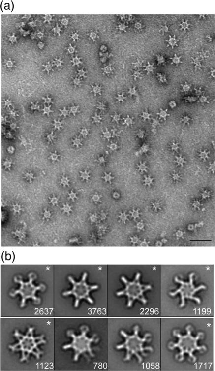
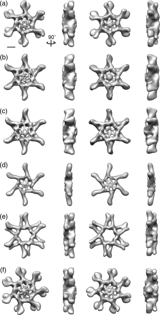
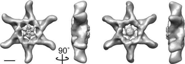
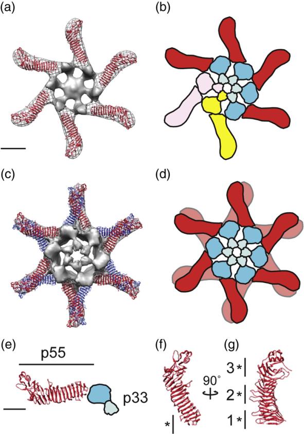
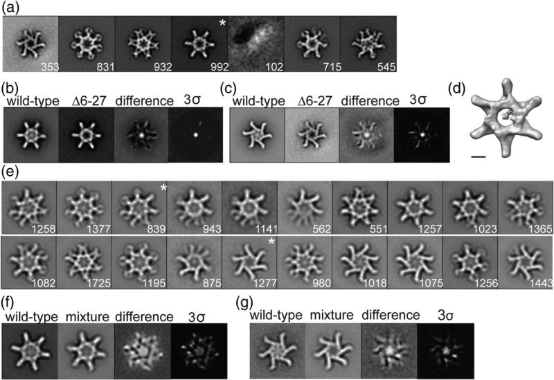
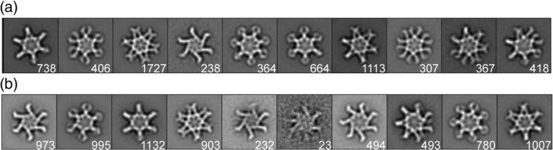
Helicobacter pylori is a Gram-negative bacterium that colonizes the human stomach and contributes to peptic ulceration and gastric adenocarcinoma. H. pylori secretes a pore-forming exotoxin known as vacuolating toxin (VacA). VacA contains two distinct domains, designated p33 and p55, and assembles into large "snowflake"-shaped oligomers. Thus far, no structural data are available for the p33 domain, which is essential for membrane channel formation. Using single-particle electron microscopy and the random conical tilt approach, we have determined the three-dimensional structures of six VacA oligomeric conformations at ~15-Å resolution. The p55 domain, composed primarily of β-helical structures, localizes to the peripheral arms, while the p33 domain consists of two globular densities that localize within the center of the complexes. By fitting the VacA p55 crystal structure into the electron microscopy densities, we have mapped inter-VacA interactions that support oligomerization. In addition, we have examined VacA variants/mutants that differ from wild-type (WT) VacA in toxin activity and/or oligomeric structural features. Oligomers formed by VacA∆6-27, a mutant that fails to form membrane channels, lack an organized p33 central core. Mixed oligomers containing both WT and VacA∆6-27 subunits also lack an organized core. Oligomers formed by a VacA s2m1 chimera (which lacks cell-vacuolating activity) and VacAΔ301-328 (which retains vacuolating activity) each contain p33 central cores similar to those of WT oligomers. By providing the most detailed view of the VacA structure to date, these data offer new insights into the toxin's channel-forming component and the intermolecular interactions that underlie oligomeric assembly.


Figures

Fig. 1
Fig. 1
Characterization of negatively stained VacA oligomers. (a) Raw image of VacA particles in negative stain. The scale bar represents 50 nm. (b) VacA class averages obtained by reference-based alignment and classification. “*” marks classes that yielded 3D structures (see Fig. 2). The number of particles included in each class is shown in the bottom right corner. Side length of panels, 420 Å.
Fig. 2
Fig. 2
3D reconstruction of negatively stained VacA oligomers using the RCT approach. (a–f) VacA organizes into a number of oligomeric conformations that include both hexamers, dodecamers, and tetradecamers. These structures have applied symmetry. Structures are rotated along the vertical axis by 90°. The scale bar represents 5 nm.
Fig. 3
Fig. 3
Approximately 15-Å 3D reconstruction of VacA dodecamer. The structure contains two prominent features: extended straight “arms” with a slight kink at the distal end and a central spoke-like density composed of two distinct globular domains separated by a thinner connecting density. The structure is rotated around the vertical axis by 90°. The scale bar represents 5 nm.
Fig. 4
Fig. 4
Structural model of the VacA oligomerization process. (a) The 2.4-Å crystal structure of p55 fits into the straight “arms” of the EM map of a VacA hexamer. Subtracting the density of the p55 crystal structure from the EM map highlights p33 (gray, central density and spokes). (b) p88 oligomerizes into hexamers supported by intermolecular interactions between the N-terminal portions of p33 in adjacent protomers, as well as contacts between p33 and an adjacent p55 arm. Blue domains, p33; red domain, p55. Two p88 protomers are colored pink and yellow to show p88 protomer interactions. (c) The 2.4-Å crystal structure of p55 fits into the straight “arms” of the EM map of a VacA dodecamer. For ease in viewing the model, the p33 density of only the well-organized side is shown. (d) Cartoon of dodecamer formation. Colors are the same as in (b). (a–d) The scale bar represents 5 nm. (e) The C-terminal p55 domain forms a straight arm with a kink at the end, while the N-terminal p33 domain consists of two globular densities connected by a thinner density (blue domains). (f and g) p55 crystal structure (2.4 Å) rotated 90° on the vertical axis. “*” marks regions of p55 involved in (f) hexamer interactions (residues 442–448) and (g) dodecamer and tetradecamer interactions. “1”, residues 395–404 and 421–435; “2”, residues 519–530 and 547–559; and “3”, residues 645–654 and 687–692. (e–g) The scale bar represents 2.5 nm.
Fig. 5
Fig. 5
Characterization of VacAΔ6–27. (a) VacAΔ6–27 class averages obtained by reference-based alignment and classification. “*”, class shown as a 3D volume in (d). (b and c) Difference mapping between WT and similar VacAΔ6–27 class averages. Final panel shown at 3 σ threshold. (a–c) Side length of panels, 573 Å. (d) 3D structure of VacAΔ6–27 corresponding to the “*” average in (a). The structure has no applied symmetry. The scale bar represents 5 nm. (e) Class averages of VacA oligomers generated by mixing WT VacA and VacAΔ6–27. “*”, classes used in difference mapping shown in (f) and (g). (f and g) Difference mapping between WT VacA and mixture of WT:VacAΔ6–27 oligomers. The final panel shown at 3 σ threshold. (e–g) Side length of panels, 420 Å. The number of particles included in each class is shown in the bottom right corner.
Fig. 6
Fig. 6
Reference-based alignment of VacAΔ301–328 and VacA s2m1. (a) VacAΔ301–328 class averages obtained by reference-based alignment and classification. (b) VacA s2m1 class averages obtained by reference-based alignment and classification. Side length of panels, 420 Å. The number of particles included in each class is shown in the bottom right corner.

References

    1. Marshall BJ, Warren JR. Unidentified curved bacilli in the stomach of patients with gastritis and peptic ulceration. Lancet. 1984;1:1311–1315. - PubMed
    1. Cover TL, Blaser MJ. Helicobacter pylori in health and disease. Gastroenterology. 2009;136:1863–1873. - PMC - PubMed
    1. Atherton JC, Blaser MJ. Coadaptation of Helicobacter pylori and humans: ancient history, modern implications. J. Clin. Invest. 2009;119:2475–2487. - PMC - PubMed
    1. Suerbaum S, Michetti P. Helicobacter pylori infection. N. Engl. J. Med. 2002;347:1175–1186. - PubMed
    1. Atherton JC. The pathogenesis of Helicobacter pylori-induced gastro-duodenal diseases. Annu. Rev. Pathol. 2006;1:63–96. - PubMed

Publication types

MeSH terms

Substances

LinkOut - more resources