. 2013 May;471(5):1639-45.
doi: 10.1007/s11999-012-2708-6. Epub 2012 Nov 21.

Does femoral rotation influence anteroposterior alpha angle, lateral center-edge angle, and medial proximal femoral angle? A pilot study

Affiliations

Does femoral rotation influence anteroposterior alpha angle, lateral center-edge angle, and medial proximal femoral angle? A pilot study

Shafagh Monazzam et al. Clin Orthop Relat Res. 2013 May.

Abstract

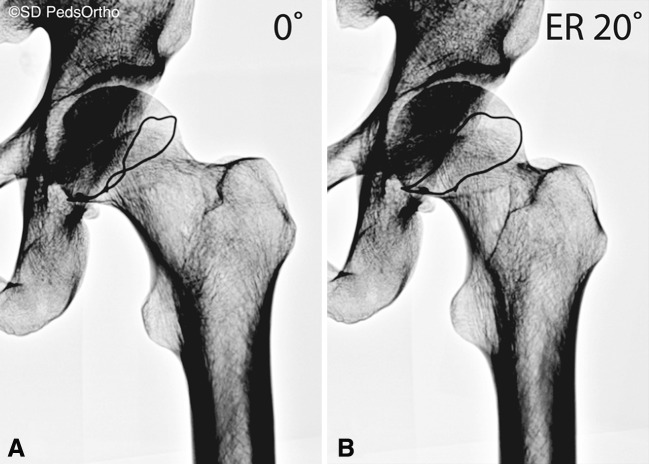
Background: Femoral rotation on AP radiographs affects several parameters used to assess morphologic features of the proximal femur but its effect on femoroacetabular impingement parameters remains unknown.

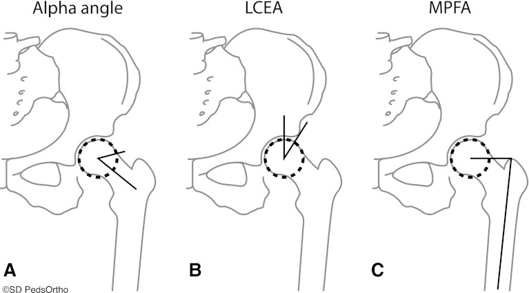
Question/purposes: We therefore evaluated and characterized the potential effect of femoral rotation on (1) AP alpha angle, (2) lateral-center edge angle (LCEA), and (3) medial proximal femoral angle (MPFA) on AP hip radiographs.

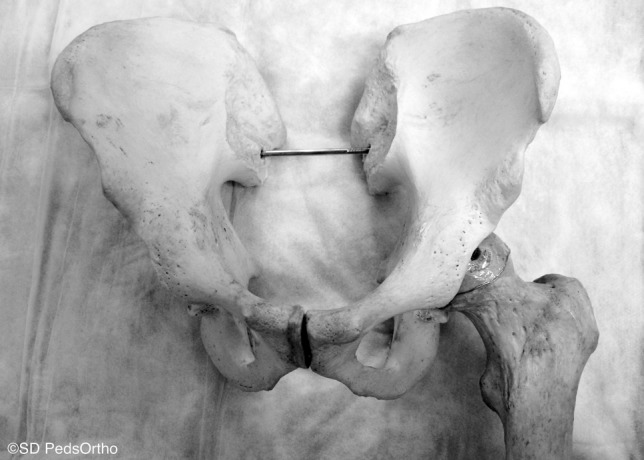
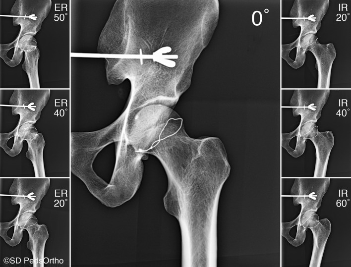
Methods: We took seven AP hip radiographs at intervals of successive femoral rotation on a single dry, cadaveric specimen: 60°, 40°, and 20° internal rotation; 0° neutral/anatomic rotation; and 20°, 40°, and 50° external rotation. The AP alpha angle, LCEA, and MPFA were measured on all radiographs by two independent evaluators.

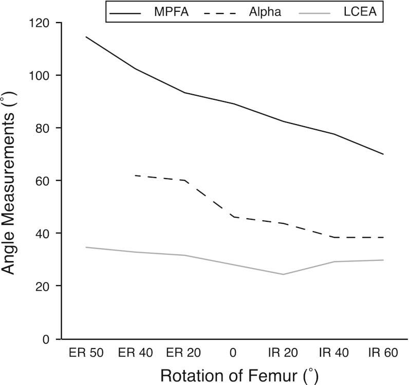
Results: Within the range of femoral rotation studied, the AP alpha angle ranged from 39° to 62°, the LCEA from 25° to 35°, and the MPFA from 70° to 115°. MPFA and AP alpha angle showed a linear relationship with femoral rotation. Each additional degree of internal rotation produced a reciprocal reduction of the MPFA by 0.36° and the AP alpha angle by 0.18° and vice versa in external rotation. The LCEA, especially within the internal rotation range, showed minimal variation.

Conclusions: These changes in radiographic parameters emphasize the importance of femoral rotation and patient positioning. We recommend radiographs be evaluated for excessive femoral rotation or nonstandardized positioning before interpretation for diagnostic and treatment implications. It may be prudent to repeat radiographs in these circumstances or, when standardized positioning is not feasible, proceed toward advance imaging.


Figures

Fig. 1
Fig. 1
The cadaveric pelvis and proximal femur used in this study are shown.
Fig. 2
Fig. 2
AP radiographs of the cadaver proximal femur in 50°, 40°, and 20° external rotation (ER), 0° neutral/anatomic rotation, and 20°, 40°, and 60° internal rotation (IR) are shown. A radiopaque wire had been glued to the cadaveric proximal femur to represent a theoretical growth plate for a previous unpublished study but was not used in this study.
Fig. 3A–C
Fig. 3A–C
The methods for measuring (A) the alpha angle, (B) lateral center-edge angle (LCEA), and (C) medial proximal femoral angle (MPFA) are shown. The alpha angle is formed between the line from the center of the femoral head to the middle of the narrowest part of the femoral neck and a line from the center of the femoral head to the point where the femoral head deviates from the circle contour. The LCEA is formed between a line from the center of the femoral head, vertically, and a line from the center of the femoral head to the lateral point of the acetabulum. The MPFA is formed between a line from the center of the femoral head to the proximal tip of the greater trochanter and a line from the proximal tip of the greater trochanter to the middle of the most distal part of the femur.
Fig. 4
Fig. 4
A graph shows the average of the two evaluators’ measurements of the alpha angle, lateral center-edge angle (LCEA), and medial proximal femoral angle (MPFA) from 50° external rotation (ER) to 60° internal rotation (IR). The effect of femoral rotation on the alpha angle and MPFA is characterized by a linear decrease in measurement with IR. The LCEA shows minimal variation with femoral rotation and no clear effect can be characterized.
Fig. 5A–B
Fig. 5A–B
AP radiographs show the cadaveric pelvis and proximal femur at (A) 0° neutral/anatomic rotation and (B) 20° external rotation (ER). No evidence of cam morphology is seen at 0° neutral/anatomic rotation, but with 20° ER, a radiograph image of cam morphology is apparent.

References

    1. Bardakos NV, Villar RN. Predictors of progression of osteoarthritis in femoroacetabular impingement: a radiological study with a minimum of ten years follow-up. J Bone Joint Surg Br. 2009;91:162–169. doi: 10.1302/0301-620X.91B2.21137. - DOI - PubMed
    1. Barton C, Salineros MJ, Rakhra KS, Beaule PE. Validity of the alpha angle measurement on plain radiographs in the evaluation of cam-type femoroacetabular impingement. Clin Orthop Relat Res. 2011;469:464–469. doi: 10.1007/s11999-010-1624-x. - DOI - PMC - PubMed
    1. Bell AL, Brand RA. Roentgenographic changes in proximal femoral dimensions due to hip rotation. Clin Orthop Relat Res. 1989;240:194–199. - PubMed
    1. Clohisy JC, Carlisle JC, Beaule PE, Kim YJ, Trousdale RT, Sierra RJ, Leunig M, Schoenecker PL, Millis MB. A systematic approach to the plain radiographic evaluation of the young adult hip. J Bone Joint Surg Am. 2008;90(suppl 4):47–66. doi: 10.2106/JBJS.H.00756. - DOI - PMC - PubMed
    1. Colvin AC, Koehler SM, Bird J. Can the change in center-edge angle during pincer trimming be reliably predicted? Clin Orthop Relat Res. 2011;469:1071–1074. doi: 10.1007/s11999-010-1581-4. - DOI - PMC - PubMed