

MDMA increases glutamate release and reduces parvalbumin-positive GABAergic cells in the dorsal hippocampus of the rat: role of cyclooxygenase
- PMID: 23179355
- PMCID: PMC3587367
- DOI: 10.1007/s11481-012-9420-x
MDMA increases glutamate release and reduces parvalbumin-positive GABAergic cells in the dorsal hippocampus of the rat: role of cyclooxygenase
Abstract
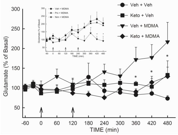
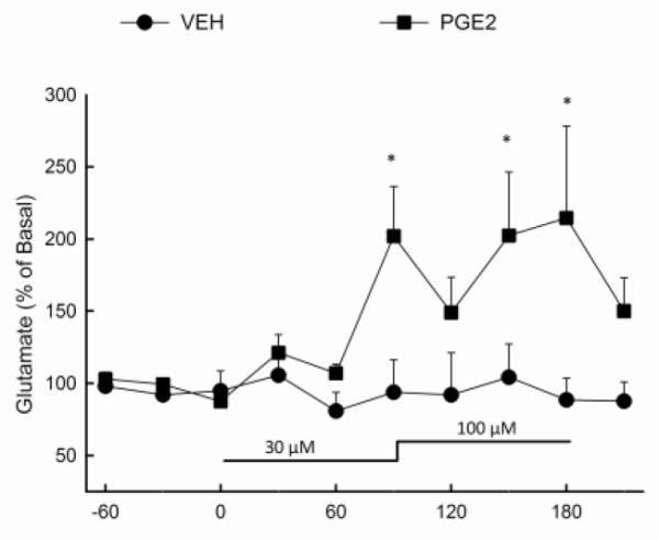
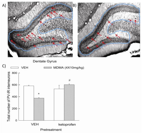
3,4-Methylenedioxymethamphetamine (MDMA; Ecstasy) is a popular drug of abuse with well-documented acute effects on serotonergic, dopaminergic, and cholinergic transmitter systems, as well as evidence of long-term disruption of serotoninergic systems in the rat brain. Recently, it was demonstrated that MDMA evokes a delayed and sustained increase in glutamate release in the hippocampus. The purpose of the present study was to determine the role of inflammatory mediators in the MDMA-induced increase in glutamate release, as well as the contribution of inflammatory pathways in the persistent neurochemical toxicity associated with repeated MDMA treatment. Treatment with the non-selective cyclooxygenase (COX) inhibitor ketoprofen and the COX-2 selective inhibitor nimesulide attenuated the increase in extracellular glutamate in the hippocampus evoked by repeated MDMA exposure (10 mg/kg, i.p., every 2 h); no attenuation was observed in rats treated with the COX-1 selective inhibitor piroxicam. Reverse dialysis of a major product of COX activity, prostaglandin E2, also resulted in a significant increase in extracellular glutamate in the hippocampus . Repeated exposure to MDMA diminished the number of parvalbumin-positive GABA interneurons in the dentate gyrus of the hippocampus, an effect that was attenuated by ketoprofen treatment. However, COX inhibition with ketoprofen did not prevent the long-term depletion of 5-HT in the hippocampus evoked by MDMA treatment. These data are supportive of the view that cyclooxygenase activity contributes to the mechanism underlying both the increased release of glutamate and decreased number of GABA interneurons in the rat hippocampus produced by repeated MDMA exposure.
Figures
References
-
- Armstrong BD, Noguchi KK. The neurotoxic effects of 3,4-methylenedioxymethamphetamine (MDMA) and methamphetamine on serotonin, dopamine, and GABA-ergic terminals: An in-vitro autoradiographic study in rats. Neurotoxicology. 2004;25:905–914. - PubMed
-
- Asanuma M, Tsuji T, Miyazaki I, Miyoshi K, Ogawa N. Methamphetamine-induced neurotoxicity in mouse brain is attenuated by ketoprofen, a non-steroidal anti-inflammatory drug. Neurosci Lett. 2003;352:13–16. - PubMed
-
- Asi SS, Farhadi HM, Mousavizadeh K, Samadikuchaksaraei A, Soleimani M, Jameie SB, Joghataei MT, Samzadeh-Kermani A, Hashemi-Nasl H, Mehdizadeh M. Evaluation of Bcl-2 Family Gene Expression in Hippocampus of 3,4-methylenedioxymethamphetamine Treated Rats. Cell Journal. 2012 2012 Winter13(4) - PMC - PubMed
Publication types
MeSH terms
Substances
Grants and funding
LinkOut - more resources
Full Text Sources
Medical
Research Materials
