

Control of cultured human cells with femtosecond laser ablated patterns on steel and plastic surfaces
- PMID: 23179464
- PMCID: PMC3595475
- DOI: 10.1007/s10544-012-9726-8
Control of cultured human cells with femtosecond laser ablated patterns on steel and plastic surfaces
Abstract
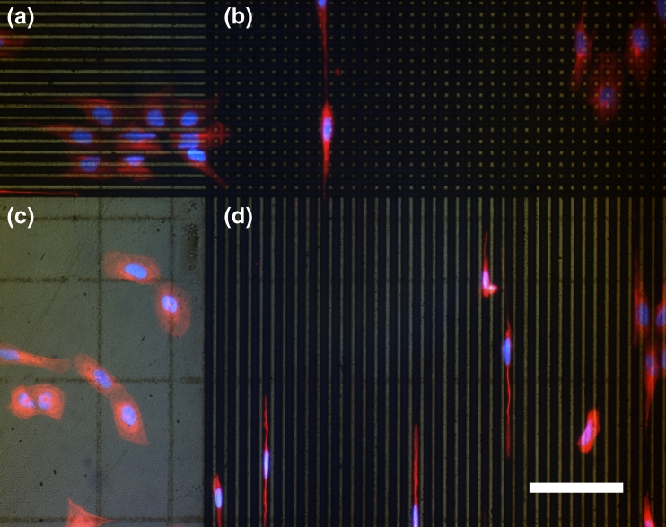
The purpose of the present study is to explore topographical patterns produced with femtosecond laser pulses as a means of controlling the behaviour of living human cells (U2OS) on stainless steel surfaces and on negative plastic imprints (polycarbonate). The results show that the patterns on both types of material strongly affect cell behaviour and are particularly powerful in controlling cell spreading/elongation, localization and orientation. Analysis by fluorescence and scanning electron microscopy shows that on periodic 1D grating structures, cells and cell nuclei are highly elongated and aligned, whereas on periodic 2D grid structures, cell spreading and shape is affected. The results also show that the density and morphology of the cells can be affected. This was observed particularly on pseudo-periodic, coral-like structures which clearly inhibited cell growth. The results suggest that these patterns could be used in a variety of applications among the fields of clinical research and implant design, as well as in diagnosis and in cell and drug research. Furthermore, this article highlights the noteworthy aspects and the unique strengths of the technique and proposes directions for further research.
Figures


Similar articles
-
In situ 24 kHz coherent imaging of morphology change in laser percussion drilling.Opt Lett. 2010 Mar 1;35(5):646-8. doi: 10.1364/OL.35.000646. Opt Lett. 2010. PMID: 20195306
-
Reduced myofibroblast differentiation on femtosecond laser treated 316LS stainless steel.Mater Sci Eng C Mater Biol Appl. 2013 Mar 1;33(2):901-8. doi: 10.1016/j.msec.2012.11.018. Epub 2012 Nov 21. Mater Sci Eng C Mater Biol Appl. 2013. PMID: 25427504
-
A facile preparation route for netlike microstructures on a stainless steel using an ethanol-mediated femtosecond laser irradiation.Mater Sci Eng C Mater Biol Appl. 2013 Mar 1;33(2):663-7. doi: 10.1016/j.msec.2012.10.014. Epub 2012 Nov 2. Mater Sci Eng C Mater Biol Appl. 2013. PMID: 25427471
-
High spatial frequency periodic structures induced on metal surface by femtosecond laser pulses.Opt Express. 2012 Jan 16;20(2):905-11. doi: 10.1364/OE.20.000905. Opt Express. 2012. PMID: 22274437
-
Micro/Nano Periodic Surface Structures and Performance of Stainless Steel Machined Using Femtosecond Lasers.Micromachines (Basel). 2022 Jun 20;13(6):976. doi: 10.3390/mi13060976. Micromachines (Basel). 2022. PMID: 35744590 Free PMC article. Review.
Cited by
-
Bone Laser Patterning to Decipher Cell Organization.Bioengineering (Basel). 2023 Jan 24;10(2):155. doi: 10.3390/bioengineering10020155. Bioengineering (Basel). 2023. PMID: 36829649 Free PMC article.
-
Effect of Laser Pulse Overlap and Scanning Line Overlap on Femtosecond Laser-Structured Ti6Al4V Surfaces.Materials (Basel). 2020 Feb 21;13(4):969. doi: 10.3390/ma13040969. Materials (Basel). 2020. PMID: 32098103 Free PMC article.
-
Synthesis of Micro-Spikes and Herringbones Structures by Femtosecond Laser Pulses on a Titanium Plate-A New Material for Water Organic Pollutants Degradation.Materials (Basel). 2021 Sep 24;14(19):5556. doi: 10.3390/ma14195556. Materials (Basel). 2021. PMID: 34639953 Free PMC article.
References
-
- Alves NM, Shi J, Oramas E, Santos JL, Tomas H, Mano JF. Bioinspired superhydrophobic poly(L-lactic acid) surfaces control bone marrow derived cells adhesion and proliferation. J. Biomed. Mater. Res. A. 2009;91:480–488. - PubMed
Publication types
MeSH terms
Substances
LinkOut - more resources
Full Text Sources
