

Stem cell dynamics in Cnidaria: are there unifying principles?
- PMID: 23179637
- PMCID: PMC7211294
- DOI: 10.1007/s00427-012-0429-1
Stem cell dynamics in Cnidaria: are there unifying principles?
Abstract
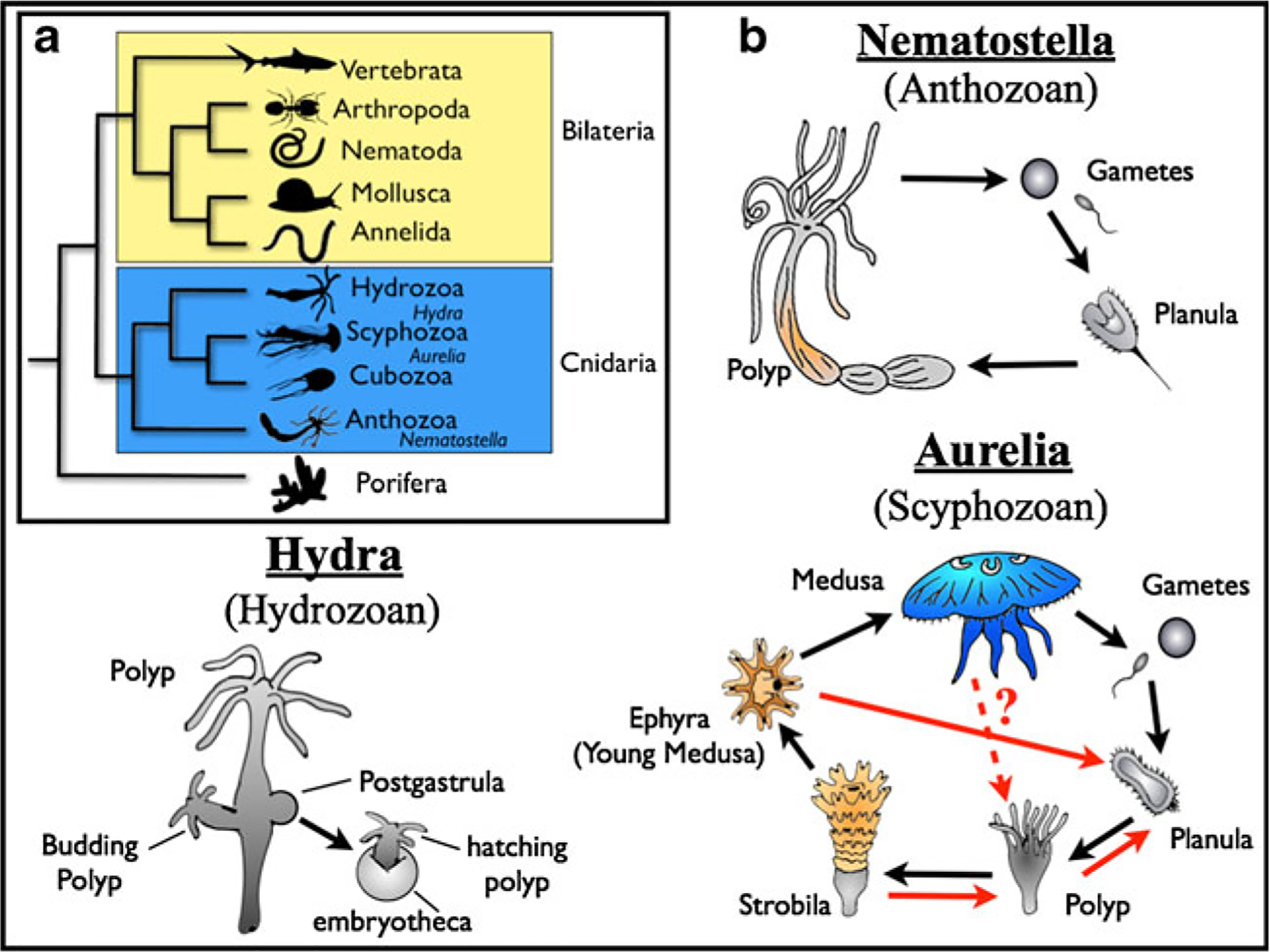
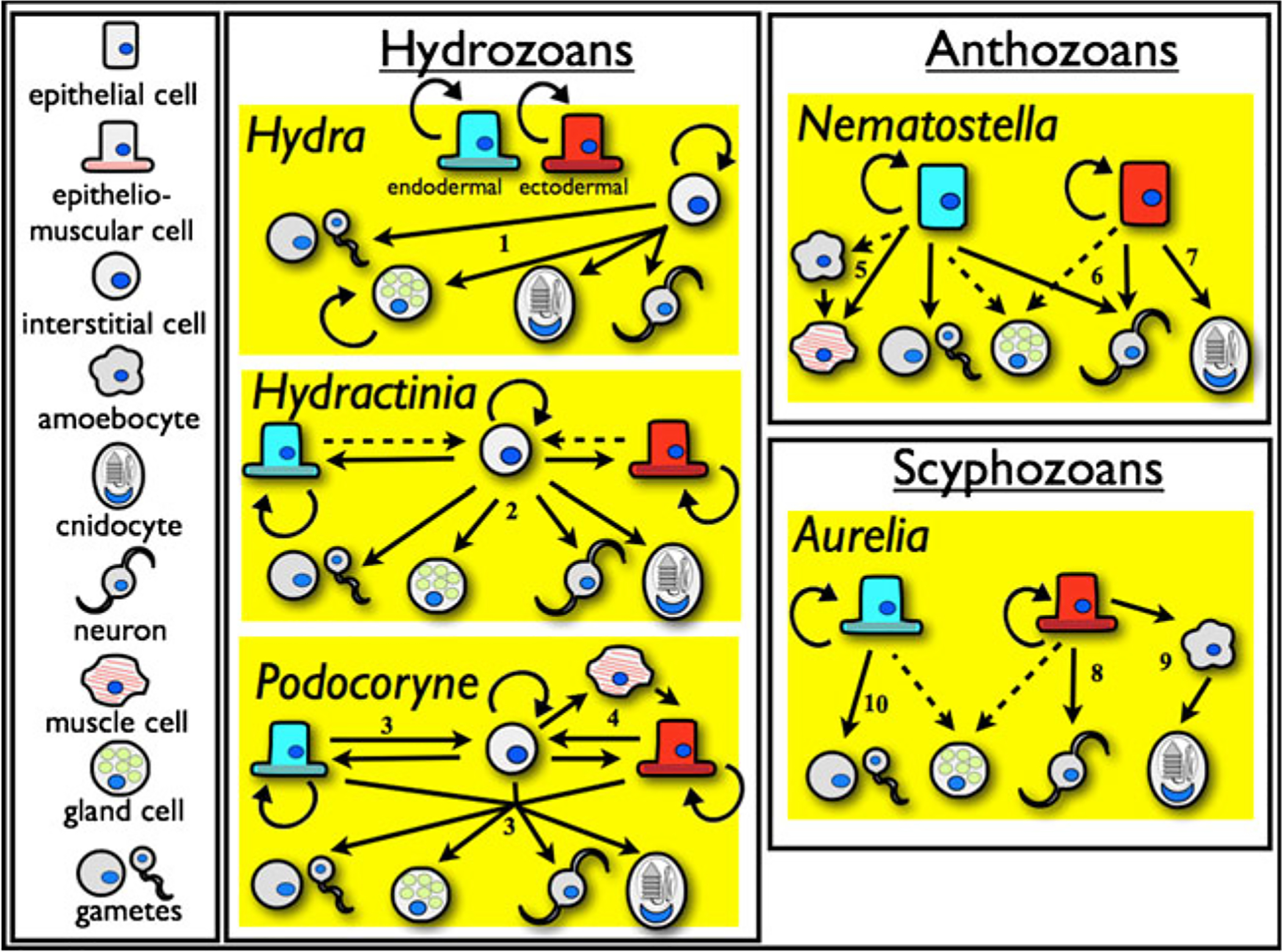
The study of stem cells in cnidarians has a history spanning hundreds of years, but it has primarily focused on the hydrozoan genus Hydra. While Hydra has a number of self-renewing cell types that act much like stem cells--in particular the interstitial cell line--finding cellular homologues outside of the Hydrozoa has been complicated by the morphological simplicity of stem cells and inconclusive gene expression data. In non-hydrozoan cnidarians, an enigmatic cell type known as the amoebocyte might play a similar role to interstitial cells, but there is little evidence that I-cells and amoebocytes are homologous. Instead, self-renewal and transdifferentiation of epithelial cells was probably more important to ancestral cnidarian development than any undifferentiated cell lineage, and only later in evolution did one or more cell types come under the regulation of a "stem" cell line. Ultimately, this hypothesis and competing ones will need to be tested by expanding genetic and developmental studies on a variety of cnidarian model systems.
Figures
Similar articles
-
Deficient autophagy in epithelial stem cells drives aging in the freshwater cnidarian Hydra.Development. 2020 Jan 23;147(2):dev177840. doi: 10.1242/dev.177840. Development. 2020. PMID: 31862842 Free PMC article.
-
Cell type complexity in the basal metazoan Hydra is maintained by both stem cell based mechanisms and transdifferentiation.Dev Biol. 2008 Jan 1;313(1):13-24. doi: 10.1016/j.ydbio.2007.09.007. Epub 2007 Sep 16. Dev Biol. 2008. PMID: 18029279
-
The Hydra polyp: nothing but an active stem cell community.Dev Growth Differ. 2010 Jan;52(1):15-25. doi: 10.1111/j.1440-169X.2009.01143.x. Epub 2009 Nov 5. Dev Growth Differ. 2010. PMID: 19891641 Review.
-
Immortality and the base of multicellular life: Lessons from cnidarian stem cells.Semin Cell Dev Biol. 2009 Dec;20(9):1114-25. doi: 10.1016/j.semcdb.2009.09.008. Epub 2009 Sep 15. Semin Cell Dev Biol. 2009. PMID: 19761866 Review.
-
Germline stem cells and sex determination in Hydra.Int J Dev Biol. 2012;56(6-8):499-508. doi: 10.1387/ijdb.123509cf. Int J Dev Biol. 2012. PMID: 22689373 Review.
Cited by
-
Harnessing the Power of Model Organisms To Unravel Microbial Functions in the Coral Holobiont.Microbiol Mol Biol Rev. 2022 Dec 21;86(4):e0005322. doi: 10.1128/mmbr.00053-22. Epub 2022 Oct 26. Microbiol Mol Biol Rev. 2022. PMID: 36287022 Free PMC article. Review.
-
Stem cells in Nanomia bijuga (Siphonophora), a colonial animal with localized growth zones.Evodevo. 2015 May 27;6:22. doi: 10.1186/s13227-015-0018-2. eCollection 2015. Evodevo. 2015. PMID: 26090088 Free PMC article.
-
The early expansion and evolutionary dynamics of POU class genes.Mol Biol Evol. 2014 Dec;31(12):3136-47. doi: 10.1093/molbev/msu243. Epub 2014 Sep 25. Mol Biol Evol. 2014. PMID: 25261405 Free PMC article.
-
Evo-devo of non-bilaterian animals.Genet Mol Biol. 2015 Jul-Sep;38(3):284-300. doi: 10.1590/S1415-475738320150005. Epub 2015 Aug 21. Genet Mol Biol. 2015. PMID: 26500432 Free PMC article.
-
A pan-metazoan concept for adult stem cells: the wobbling Penrose landscape.Biol Rev Camb Philos Soc. 2022 Feb;97(1):299-325. doi: 10.1111/brv.12801. Epub 2021 Oct 6. Biol Rev Camb Philos Soc. 2022. PMID: 34617397 Free PMC article.
References
-
- Afzelius B, Rosen B (1965) Nutritive phagocytosis in animal cells—an electron microscopical study of gastroderm of hydroid Clava squamata Mull. Z Zellforsch Mikrosk Anat 67:24. - PubMed
-
- Alié A, Leclère L, Jager M, Dayraud C, Chang P, Le Guyader H, Queinnec E, Manuel M (2011) Somatic stem cells express Piwi and Vasa genes in an adult ctenophore: ancient association of “germline genes” with stemness. Dev Biol 350:183–197 - PubMed
-
- Barnes R, Harrison F (1990) Introduction In: Harrison F, Westfall J (eds) Microscopic anatomy of invertebrates. Volume 2: Placozoa, Porifera, Cnidaria, and Ctenophora, 1st edn Wiley-Liss, New York
-
- Black RE, Riley GK (1985) Dissociation and reaggregation of cells of Chrysaora quinquecirrha (Cnidaria, Scyphozoa). J Exp Zool 233:369–375 - PubMed
Publication types
MeSH terms
Grants and funding
LinkOut - more resources
Full Text Sources
Medical
