. 2013 May;34(5):1091-7.
doi: 10.3174/ajnr.A3341. Epub 2012 Nov 22.

Imaging characteristics of cerebrovascular arteriopathy and stroke in Hutchinson-Gilford progeria syndrome

Affiliations

Imaging characteristics of cerebrovascular arteriopathy and stroke in Hutchinson-Gilford progeria syndrome

V M Silvera et al. AJNR Am J Neuroradiol. 2013 May.

Abstract

Background and purpose: HGPS is a rare disorder of segmental aging, with early morbidity from cardiovascular and cerebrovascular disease. The goal of this study was to identify the neurovascular features, infarct type, topography, and natural history of stroke in the only neurovascular imaging cohort study of HGPS.

Materials and methods: We studied 25 children with confirmed diagnoses of HGPS and neuroimaging studies available for review. Relevant clinical information was abstracted from medical records.

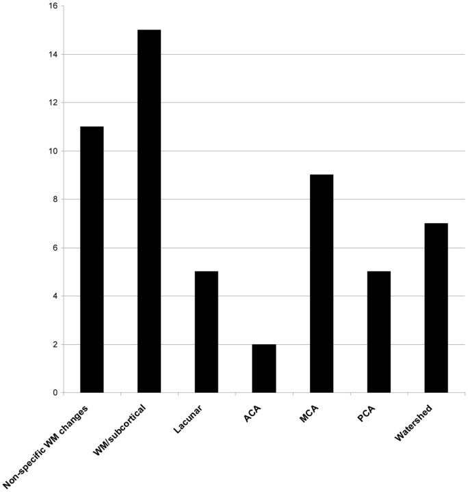
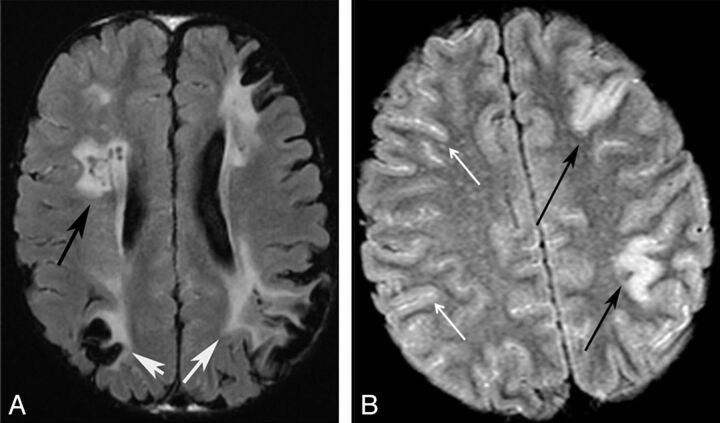
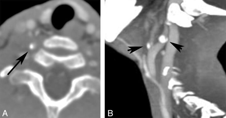
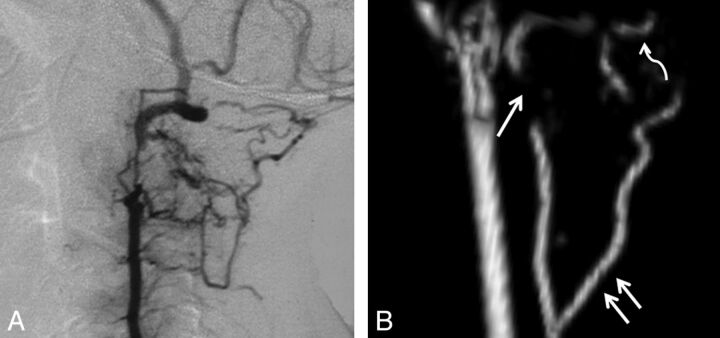
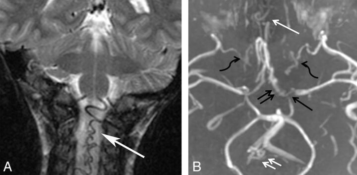
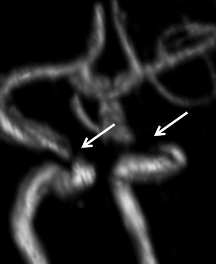
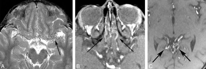
Results: We identified features suggestive of a vasculopathy unique to HGPS, including distinctive intracranial steno-occlusive arterial lesions, basal cistern collateral vessels, and slow compensatory collateral flow over the cerebral convexities. The arterial pathology in the neck consisted of distal vertebral artery stenosis with prominent collateral vessel formation as well as stenosis and calcification of both the cervical internal and common carotid arteries. Radiographic evidence of infarction was found in 60% of patients, of which half were likely clinically silent. Both large- and small-vessel disease was observed, characterized by arterial territorial, white matter, lacunar, and watershed infarcts.

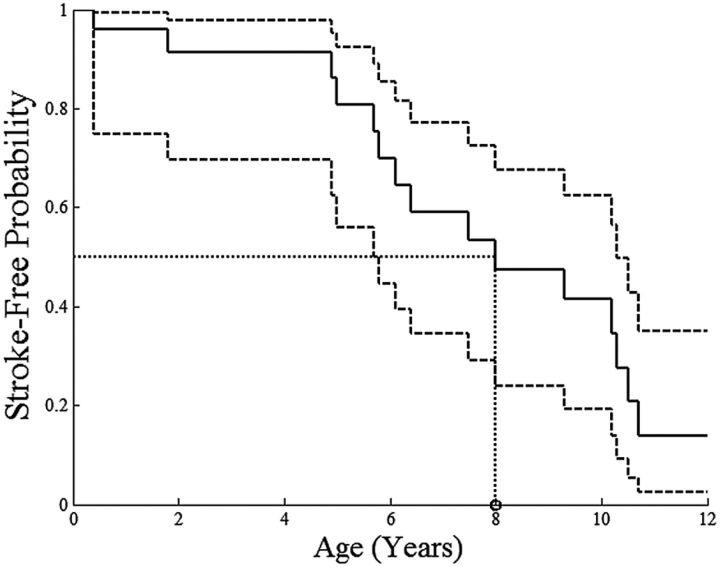
Conclusions: We report a unique intracranial and superior cervical arteriopathy in HGPS distinct from other vasculopathies of childhood, such as Moyamoya, and cerebrovascular disease of aging, including atherosclerosis. Arterial features of the mid and lower neck are less distinctive. For the first time, we identified early and clinically silent strokes as a prevalent disease characteristic in HGPS. Longitudinal analysis of stroke incidence and vasculopathy may provide an outcome measure for future treatment interventions for children with HGPS.


Figures

Fig 1.
Fig 1.
Kaplan-Meier model of stroke-free probability depicts the youngest ages of patients at which imaging demonstrated ≥1 infarct, acute or chronic. The solid line shows the estimated stroke-free probability; dashed lines, the 95% CIs; and the dotted lines, the estimated age at which 50% of children have radiographically detectable stroke.
Fig 2.
Fig 2.
Graphic representation of infarct distribution.
Fig 3.
Fig 3.
Patterns of infarction. Axial FLAIR images of 2 different patients. A, Chronic watershed (white arrows) and white matter infarcts (black arrow). B, Acute gyral infarcts (black arrows). Bright signal in the sulci indicates slow cortical collateral flow (white arrows).
Fig 4.
Fig 4.
Arterial calcification. CTA reformatted images of the same patient demonstrate right VA calcification (A) and ICA and external carotid artery (B) calcifications.
Fig 5.
Fig 5.
VA stenoses and collaterals. A, Lateral projection of a conventional angiogram demonstrates a short-segment high-grade stenosis of the distal V2 segment of the VA. B, Lateral projection of a 3D model of the cervical MRA in a different patient demonstrates a high-grade stenosis of the V2 segment of the VA (single white arrow), an enlarged deep cervical artery (double white arrows), and enlargement of the occipital artery (curved white arrow).
Fig 6.
Fig 6.
Collateral vessels. A, Coronal T2WI of an enlarged ASA (white arrow). B, Collapsed view of an MRA in a different patient demonstrates M1 (single black arrow) and A1 stenoses (black double arrows), internal maxillary artery collaterals (black wavy arrows), subfrontal collaterals (single white arrow), and an enlarged ASA and posterior spinal artery (white double arrows).
Fig 7.
Fig 7.
ICA stenosis. 3D model of the MRA of the circle of Willis demonstrates bilateral short-segment high-grade stenoses of the cavernous ICAs (white arrows).
Fig 8.
Fig 8.
Collateral vessels. A, Axial T2WI shows collateral vessels in the basal cistern (arrow). Source images from an MRA in a different patient demonstrate subfrontal (B) and perisplenial (C) collateral vessels (arrows.)

References

    1. Hennekam RC. Hutchinson-Gilford progeria syndrome: review of the phenotype. Am J Med Genet A 2006;140:2603–24 - PubMed
    1. Eriksson M, Brown WT, Gordon LB, et al. . Recurrent de novo point mutations in lamin A cause Hutchinson-Gilford progeria syndrome. Nature 2003;423:293–98 - PMC - PubMed
    1. De Sandre-Giovannoli A, Bernard R, Cau P, et al. . Lamin A truncation in Hutchinson-Gilford progeria. Science 2003;300:2055. - PubMed
    1. Goldman RD, Shumaker DK, Erdos MR, et al. . Accumulation of mutant lamin A causes progressive changes in nuclear architecture in Hutchinson-Gilford progeria syndrome. Proc Natl Acad Sci U S A 2004;101:8963–68 - PMC - PubMed
    1. Varga R, Eriksson M, Erdos MR, et al. . Progressive vascular smooth muscle cell defects in a mouse model of Hutchinson-Gilford progeria syndrome. Proc Natl Acad Sci U S A 2006;103:3250–55 - PMC - PubMed

Publication types