Review
. 2013 May;91(5):549-59.
doi: 10.1007/s00109-012-0983-z. Epub 2012 Nov 22.

Molecular targets for treatment of kidney fibrosis

Affiliations
Review

Molecular targets for treatment of kidney fibrosis

Peter Y Chuang et al. J Mol Med (Berl). 2013 May.

Abstract

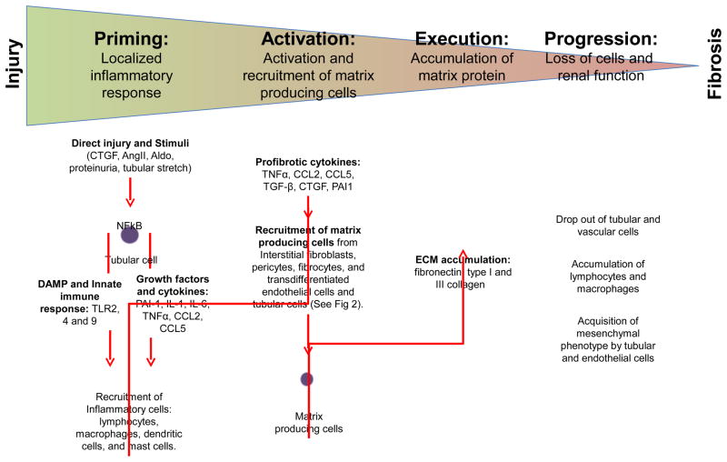
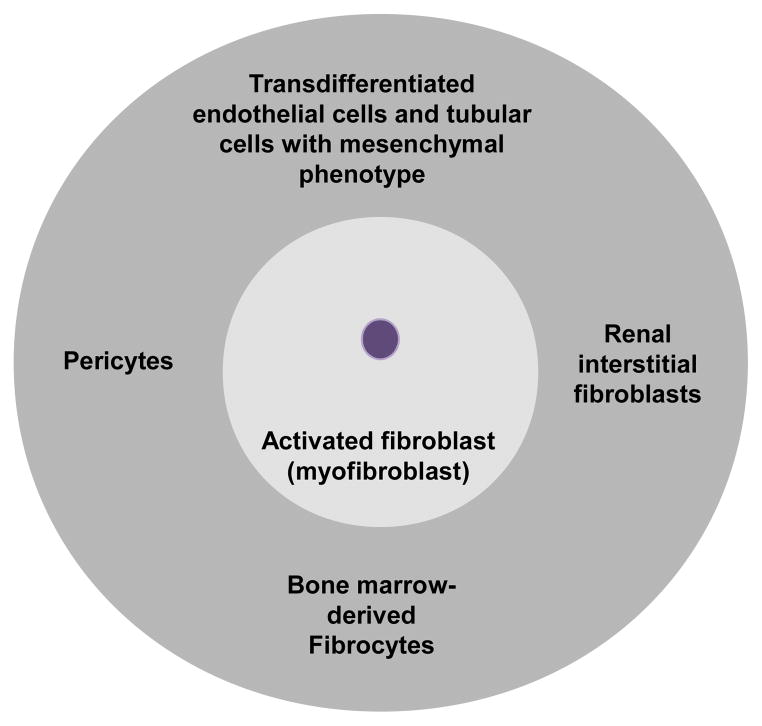
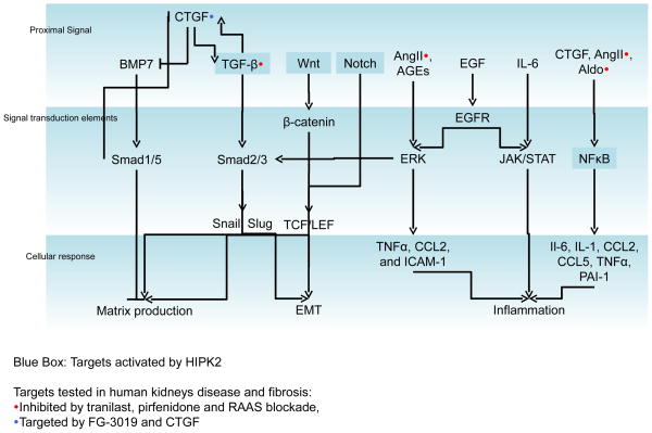
Renal fibrosis is the culmination of processes driven by signaling pathways involving transforming growth factor-β family of cytokines, connective-tissue growth factor, nuclear factor κB, Wnt/β-catenin, Notch, and other growth factors. Many studies in experimental animal models have directly targeted these pathways and demonstrated efficacy in mitigating renal fibrosis. However, only a small fraction of these approaches have been attempted in human and even fewer have been successfully translated to clinical use for patient with kidney diseases. Drugs with proven efficacy for treatment of kidney diseases and tissue fibrosis exert some of their effects by interfering with components of these pathways. This review considers key molecular mediators of renal fibrosis and their potential as targets for treatment of renal fibrosis.


Figures

Figure 1
Figure 1
Four overlapping phases of renal fibrosis: priming, activation, execution, and progression. Direct tubular epithelial cell injury or cellular stimuli triggers a pro-inflammatory response involving activation of the innate immune response and production of growth factors and cytokines, which result in the recruitment of inflammatory cells. Localized accumulation of profibrotic cytokines promotes activation and recruitment of matrix-producing cells from different sources. Accumulation of extracellular matrix proteins is observed in renal fibrosis in conjunction with loss of tubular and vascular cells, accumulation of lymphocytes and macrophages, and acquisition of mesenchymal cellular phenotype by tubular and endothelial cells, which are associated with loss of kidney function. CTGF: connective tissue growth factor, AngII: angiotensin II, Aldo: aldosterone, AGEs: advanced glycation endproducts, NFκB: nuclear factor kappa B, TLR: toll-like receptors, DAMP: danger associated molecular pattern molecules, ROS: reactive oxygen species, IL: interleukin, TGF: transforming growth factor, TNF: tumor necrosis factor, CCL: chemokine C-C motif ligand, PAI: plasminogen activator inhibitor, and ECM: extracellular matrix.
Figure 2
Figure 2
Multiple origins of myofibroblasts in renal fibrosis. Renal tubular interstitial fibroblasts, bone-marrow-derived fibrocytes, vascular pericytes, and transdifferentiated endothelial cells and tubular cells with mesenchymal phenotype have been shown to contribute to the population of myofibroblasts in the fibrotic kidney.
Figure 3
Figure 3
Schematic of key mediators of kidney fibrosis. Proximal signals from growth factors, cytokines and signaling molecules activate cellular signal transduction elements in tubular cells as well as other cell types to promote profibrotic cellular responses that include extracellular matrix production, epithelial-mesenchymal transdifferentiation, and inflammation. Signaling transduction elements are activated by different proximal signals. Homeo-domain interacting protein kinase (HIPK) 2 interfaces with nuclear factor (NF)-κB, Wnt/β-catenin, and Notch signaling components to promote renal fibrosis (highlighted in blue boxes). Approaches to antagonize CTGF, TGF-β, and renin-angiotensin-aldosterone signaling in renal disease and fibrosis have demonstrated clinical efficacy.

References

    1. Harris RC, Neilson EG. Toward a unified theory of renal progression. Annual review of medicine. 2006;57:365–380. - PubMed
    1. Striker GE, Schainuck LI, Cutler RE, Benditt EP. Structural-functional correlations in renal disease. I. A method for assaying and classifying histopathologic changes in renal disease. Human pathology. 1970;1:615–630. - PubMed
    1. Nath KA. Tubulointerstitial changes as a major determinant in the progression of renal damage. American journal of kidney diseases : the official journal of the National Kidney Foundation. 1992;20:1–17. - PubMed
    1. Mackensen-Haen S, Bader R, Grund KE, Bohle A. Correlations between renal cortical interstitial fibrosis, atrophy of the proximal tubules and impairment of the glomerular filtration rate. Clinical nephrology. 1981;15:167–171. - PubMed
    1. Boor P, Sebekova K, Ostendorf T, Floege J. Treatment targets in renal fibrosis. Nephrology, dialysis, transplantation : official publication of the European Dialysis and Transplant Association - European Renal Association. 2007;22:3391–3407. - PubMed

Publication types

MeSH terms

LinkOut - more resources