. 2013 Feb;22(2):411-6.
doi: 10.1007/s00586-012-2585-4. Epub 2012 Nov 20.

The effect of posterior spinal fusion on respiratory function in Duchenne muscular dystrophy

Affiliations

The effect of posterior spinal fusion on respiratory function in Duchenne muscular dystrophy

W M Alexander et al. Eur Spine J. 2013 Feb.

Abstract

Purpose: Posterior instrumented spinal fusion is indicated for progressive scoliosis that develops in Duchenne muscular dystrophy (DMD) patients. Whilst spinal fusion is known to improve quality of life, there is inconsistency amongst the literature regarding its specific effect on respiratory function. Our objective was to determine the effect of scoliosis correction by posterior spinal fusion on respiratory function in a large cohort of patients with DMD. Patients with DMD undergoing posterior spinal fusion were compared to patients with DMD not undergoing surgical intervention.

Methods: An observational study of 65 patients with DMD associated scoliosis, born between 1961 and 2001: 28 of which underwent correction of scoliosis via posterior spinal fusion (Surgical Group) and 37 of which did not undergo surgical intervention (Non-Surgical Group). Pulmonary function was assessed using traditional spirometry. Comparisons were made between groups at set times, and by way of rates of change over time.

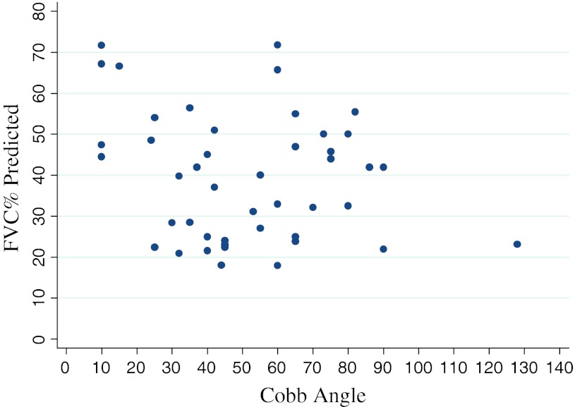
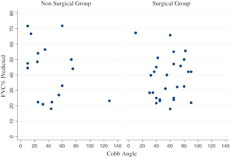
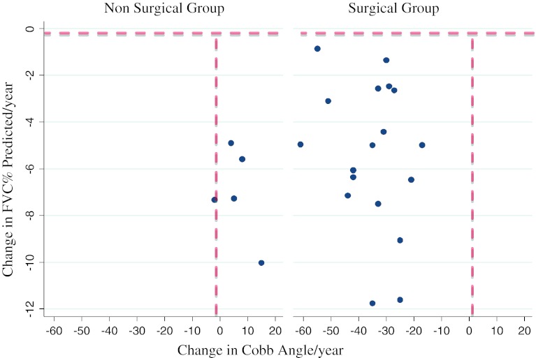
Results: There was no correlation between the level of respiratory dysfunction and the severity of scoliosis (as measured by Cobb angle) for the whole cohort. The Surgical Group had significantly worse respiratory function at a comparable age pre-operatively compared to the Non-Surgical Group, as measured by per cent predicted forced vital capacity (p = 0.02) on spirometry. The rate of decline of forced vital capacity and per cent predicted forced vital capacity was not slowed following surgery compared to the non-operated cases. There was no significant difference in survival between the two groups.

Conclusions: Severity of scoliosis was not a key determinant of respiratory dysfunction. Posterior spinal fusion did not reduce the rate of respiratory function decline. These two points suggest that intrinsic respiratory muscle weakness is the main determinant of decline in respiratory function in DMD.


Figures

Fig. 1
Fig. 1
Relationship between Cobb angle and % predicted FVC for the entire cohort (both groups) at 1st time point. No correlation was observed between the severity of scoliosis (Cobb angle) and level of respiratory dysfunction (FVC %) (p = 0.16)
Fig. 2
Fig. 2
Relationship between Cobb angle and % predicted FVC for the Non-Surgical Group (NSG) and the Surgical Group (SG). No correlation was observed between the severity of scoliosis (Cobb angle) and level of respiratory dysfunction (FVC %) in either group (NSG: p = 0.19; SG: p = 0.96)
Fig. 3
Fig. 3
Change in Cobb angle versus Change in % predicted FVC in Non-Surgical Group (NSG) (left) and Surgical Group (SG) (right). Both graphs depict change in Cobb angle, which can be either negative (indicating a improvement in scoliosis) or positive (indicating worsening scoliosis). The vertical dashed lines on both graphs represents no change. Note that all the NSG (left) had INCREASES in the Cobb angle; whereas all the SG (right) had decreases in Cobb angles in the range 20°–60°. The Y-axis shows change in FVC. Note the horizontal dashed line represents zero change and indicates that all patients deteriorated in respiratory function between the two time points. This shows that regardless of whether the Cobb angle deteriorates in the NSG, or improves in the SG, the FVC continues to decline. Along with the previous graph, this indicates that there is no association between Cobb angles and FVCs, even when measured over time with significant improvement in the Cobb angle (NSG: p = 0.37; SG: p = 0.34). Outliers have been excluded

Similar articles

Cited by

References

    1. Hoffman EP, Fischbeck KH. Characterization of dystrophin in muscle-biopsy specimens from patients with Duchenne’s or Becker’s muscular dystrophy. N Engl J Med. 1988;318(21):1363–1368. doi: 10.1056/NEJM198805263182104. - DOI - PubMed
    1. Marsh A, Edge G, Lehovsky J. Spinal fusion in patients with Duchenne’s muscular dystrophy and a low forced vital capacity. Eur Spine J. 2003;12(5):507–512. doi: 10.1007/s00586-003-0545-8. - DOI - PMC - PubMed
    1. Kennedy JD, Staples AJ, Brook PD, et al. Effect of spinal surgery on lung function in Duchenne muscular dystrophy. Thorax. 1995;50(11):1173–1178. doi: 10.1136/thx.50.11.1173. - DOI - PMC - PubMed
    1. Emery A, Muntoni F. Duchenne muscular dystrophy. Oxford: Oxford University Press; 2003.
    1. Eagle M, Bourke J, Bullock R, et al. Managing Duchenne muscular dystrophy–the additive effect of spinal surgery and home nocturnal ventilation in improving survival. Neuromuscul Disord. 2007;17(6):470–475. doi: 10.1016/j.nmd.2007.03.002. - DOI - PubMed