. 2013 Jan;55(1):33-46.
doi: 10.1007/s10858-012-9683-9. Epub 2012 Nov 23.

Database proton NMR chemical shifts for RNA signal assignment and validation

Affiliations

Database proton NMR chemical shifts for RNA signal assignment and validation

Shawn Barton et al. J Biomol NMR. 2013 Jan.

Abstract

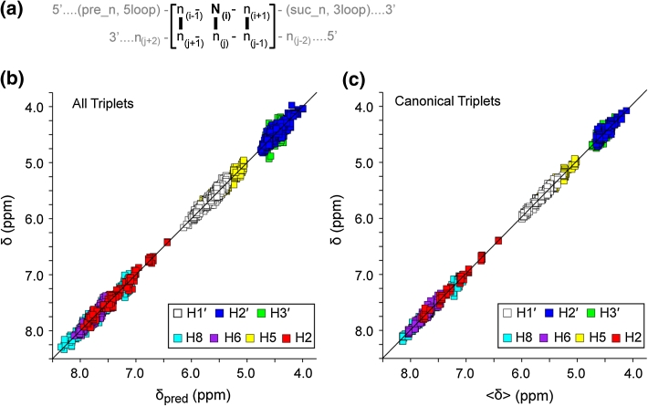
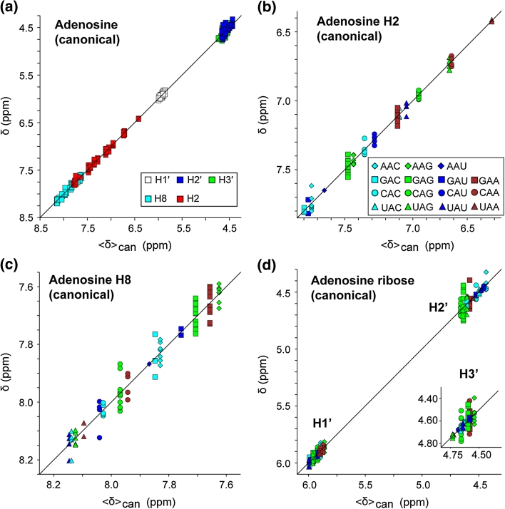
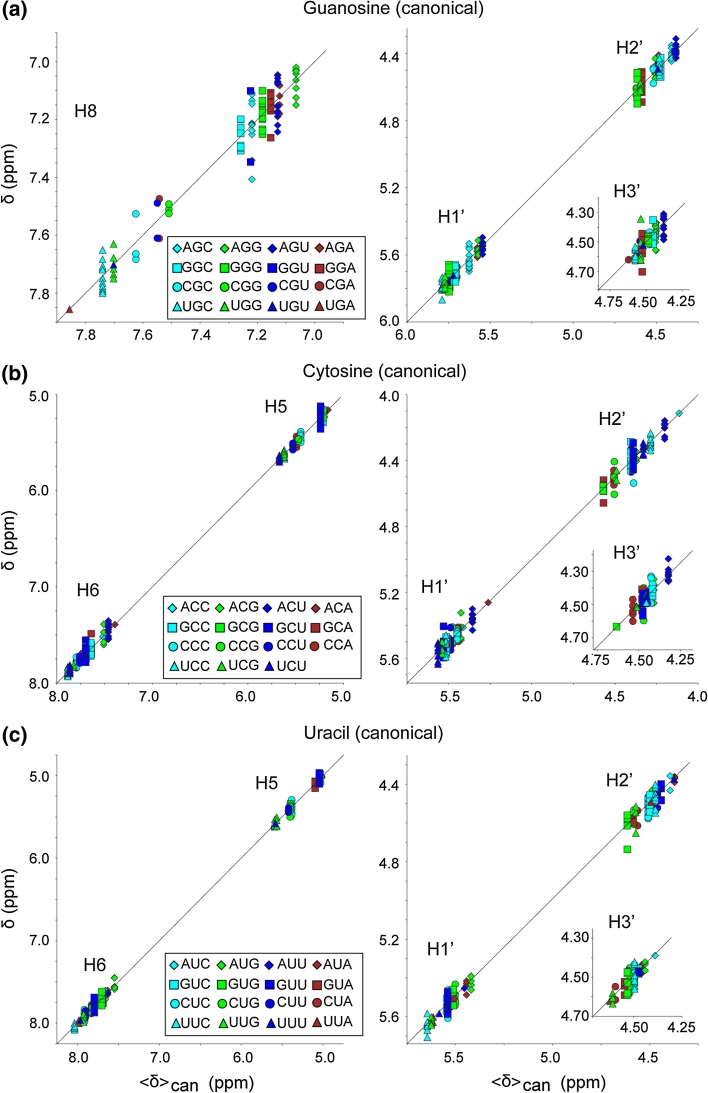
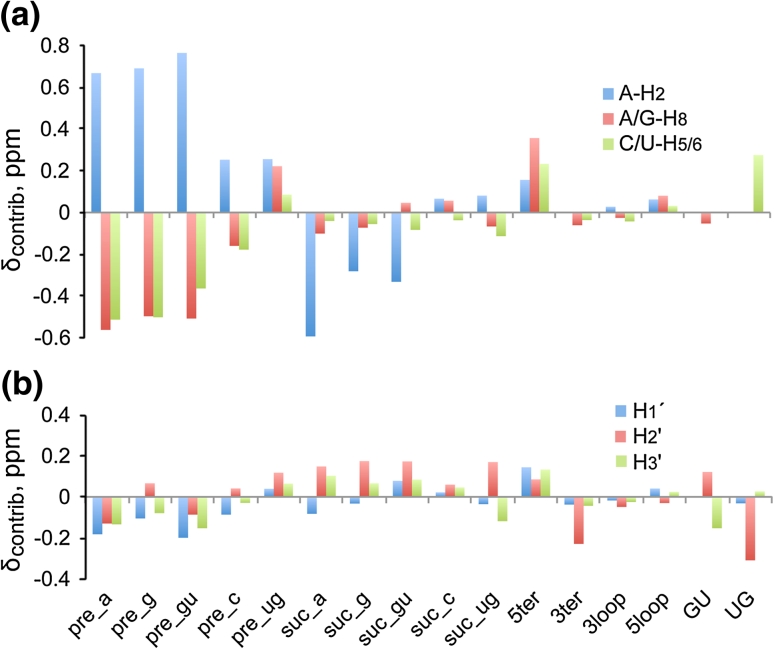
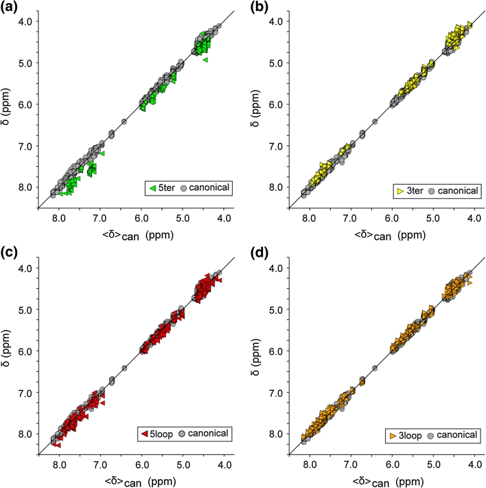
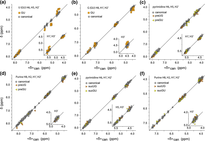
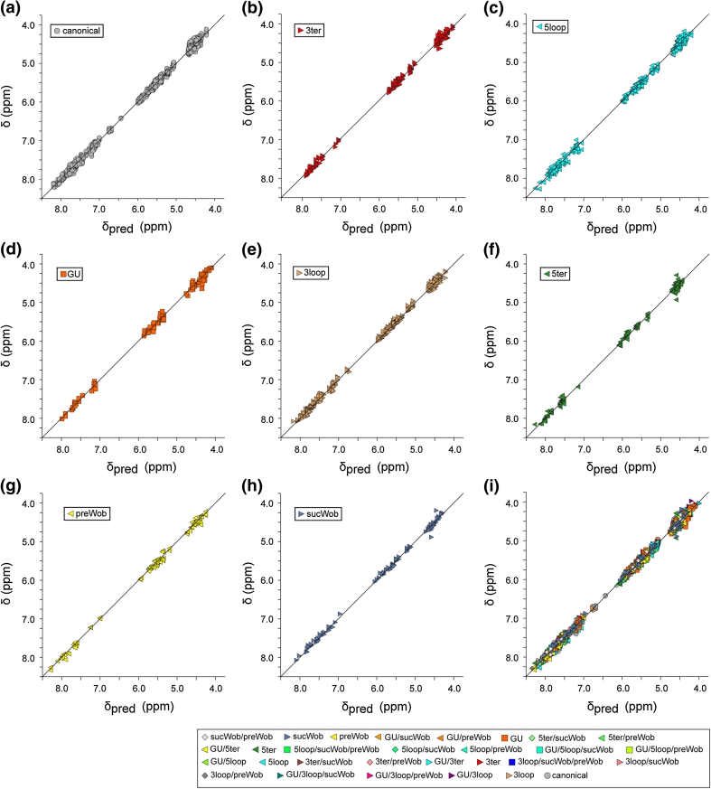
The Biological Magnetic Resonance Data Bank contains NMR chemical shift depositions for 132 RNAs and RNA-containing complexes. We have analyzed the (1)H NMR chemical shifts reported for non-exchangeable protons of residues that reside within A-form helical regions of these RNAs. The analysis focused on the central base pair within a stretch of three adjacent base pairs (BP triplets), and included both Watson-Crick (WC; G:C, A:U) and G:U wobble pairs. Chemical shift values were included for all 4(3) possible WC-BP triplets, as well as 137 additional triplets that contain one or more G:U wobbles. Sequence-dependent chemical shift correlations were identified, including correlations involving terminating base pairs within the triplets and canonical and non-canonical structures adjacent to the BP triplets (i.e. bulges, loops, WC and non-WC BPs), despite the fact that the NMR data were obtained under different conditions of pH, buffer, ionic strength, and temperature. A computer program (RNAShifts) was developed that enables convenient comparison of RNA (1)H NMR assignments with database predictions, which should facilitate future signal assignment/validation efforts and enable rapid identification of non-canonical RNA structures and RNA-ligand/protein interaction sites.


Figures

Fig. 1
Fig. 1
a Definitions used for base pair triplets. The chemical shifts of the N(i) residue are analyzed in this work, and this strand may be preceded by a base-paired (WC or GU wobble) nucleotide (pre_n) or a non-base paired residue (5loop), or followed by a base-paired residue (suc_n) or non-base paired residue (3loop). b Plot of the database chemical shift (automatically re-referenced as described in the text) (δ) versus calculated chemical shift (δpred) for the 3758 assignment depositions utilized in the present study (rms deviation = 0.056). c Plot of δ versus mean chemical shift (〈δ〉) for residues in canonical triplets (triplets that contain only GC and/or AU base pairs and are both preceded and followed by a GC and/or AU base pair) (rms deviation = 0.043)
Fig. 2
Fig. 2
Plots of re-referenced 1H NMR chemical shifts (δ) reported for the central adenosine residues within canonical triplets (as defined in text and Fig. 1 caption) versus mean shifts calculated for canonical triplets (〈δ〉can). a Data are shown for all adenosine protons grouped by atom type (symbols defined in a inset). bd Expansions showing data grouped according to triplet sequence for the adenosine H2 (b), H8 (c) and ribose (d) protons (symbols defined in b inset)
Fig. 3
Fig. 3
Plots of re-referenced 1H NMR chemical shifts (δ) reported for the central guanosine (a), cytosine (b) and uracil (c) residues within canonical triplets (as defined in text and Fig. 1) versus mean shifts calculated for canonical triplets (〈δ〉can). Data are grouped by atom type as defined in panel insets
Fig. 4
Fig. 4
Plot of the chemical shift contributions (δcontrib) of each attribute relative to a canonical uNu triplet as obtained via Pace Regression for aromatic (a) and ribose (b) proton assignments (positive values denote downfield shifts). Data in these plots are derived from Table 1. For simplification, data for aromatic protons with similar trends in their response to the attributes were combined, and within each group of proton type, the largest absolute value is plotted. Because this procedure can mask the details of individual proton types one should use this plot for observing general trends and refer to the specific contributions in Table 1
Fig. 5
Fig. 5
Plots of δ versus 〈δ〉can (defined in Figs. 1, 2 captions) for the central residues of WC-BP triplets that contain a 5′-terminal base pair (5ter), a 3′-terminal base pair (3ter), or are preceded and/or followed by non-canonical loops or bulges (5loop and 3loop, respectively). Symbols are defined in the panel insets
Fig. 6
Fig. 6
Plots showing the sensitivity of the 1H NMR chemical shifts to GU and UG wobble pairing within the canonical WC-BP triplet. a The central U of an otherwise canonical triplet is paired with G. b The central G of an otherwise canonical triplet is paired with U. c–f Influence of GU base pairs at the n(i−1) and n(i+1) position of the n(i−1)−Ni−n(i+1) triplet on the NMR chemical shift of the central canonical base pair. Symbols are defined in the panel insets
Fig. 7
Fig. 7
Plots of δ versus predicted chemical shift (δpred), calculated by Pace regression as described in the text. ah Data for triplets that are fully canonical (a) (rmsd = 0.050) or include a single non-canonical element, b 3′-terminal residue (3ter), rmsd = 0.054; c loop, bulge or stacked non-BP residue immediately 5′ to the triplet, rmsd = 0.066; d GU wobble at the center of an otherwise canonical triplet (GU), rmsd = 0.073; e loop, bulge or stacked non-BP residue immediately 3′ to the triplet (3ter), rmsd = 0.053; f 5′-terminal residue within the triplet (5ter), rmsd = 0.054; g 5′-residue of the triplet is involved in a GU wobble interaction (preWob), rmsd = 0.057; h 3′-residue of the triplet is involved in a GU wobble interaction (sucWob), rmsd = 0.057; i all data, including triplets with multiple non-canonical elements, rmsd = 0.056

References

    1. Aeschbacher T, Schubert M, Allain FH. A procedure to validate and correct the 13C chemical shift calibration of RNA datasets. J Biomol NMR. 2012;52:179–190. doi: 10.1007/s10858-011-9600-7. - DOI - PubMed
    1. Allain FH-T, Varani G. How accurately and precisely can RNA structure be determined by NMR? J Mol Biol. 1997;267:338–351. doi: 10.1006/jmbi.1996.0855. - DOI - PubMed
    1. Bahrami A, Clos LJ, 2nd, Markley JL, Butcher SE, Eghbalnia HR. RNA-PAIRS: RNA probabilistic assignment of imino resonance shifts. J Biomol NMR. 2012;52:289–302. doi: 10.1007/s10858-012-9603-z. - DOI - PMC - PubMed
    1. Bartel DP. MicroRNAs: genomics, biogenesis, mechanism, and function. Cell. 2004;116:281–297. doi: 10.1016/S0092-8674(04)00045-5. - DOI - PubMed
    1. Batey RT, Inada M, Kujawinski E, Puglisi JD, Williamson JR. Preparation of isotopically labeled ribonucleotides for multidimensional NMR spectroscopy of RNA. Nucleic Acids Res. 1992;20:4515–4523. doi: 10.1093/nar/20.17.4515. - DOI - PMC - PubMed

Publication types

LinkOut - more resources