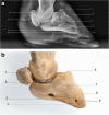
The radiographic anatomy of the normal ovine digit, the metacarpophalangeal and metatarsophalangeal joints
- PMID: 23180463
- PMCID: PMC3565091
- DOI: 10.1007/s11259-012-9546-6
The radiographic anatomy of the normal ovine digit, the metacarpophalangeal and metatarsophalangeal joints
Abstract
The aim of this project was to develop a detailed, accessible set of reference images of the normal radiographic anatomy of the ovine digit up to and including the metacarpo/metatatarsophalangeal joints. The lower front and hind limbs of 5 Lleyn ewes were radiographed using portable radiography equipment, a digital image processer and standard projections. Twenty images, illustrating the normal radiographic anatomy of the limb were selected, labelled and presented along with a detailed description and corresponding images of the bony skeleton. These images are aimed to be of assistance to veterinary surgeons, veterinary students and veterinary researchers by enabling understanding of the normal anatomy of the ovine lower limb, and allowing comparison with the abnormal.
Figures


References
-
- FAWC (2011) Opinion on lameness in sheep. Farm Animal Welfare Council.
-
- Hodgkinson O. The importance of feet examination in sheep health management. Small Ruminant Res. 2010;92:67–71. doi: 10.1016/j.smallrumres.2010.04.007. - DOI
MeSH terms
LinkOut - more resources
Full Text Sources

