. 2013 Jan 4;339(6115):70-4.
doi: 10.1126/science.1227622. Epub 2012 Nov 22.

The spatial and temporal origin of chandelier cells in mouse neocortex

Affiliations

The spatial and temporal origin of chandelier cells in mouse neocortex

Hiroki Taniguchi et al. Science. .

Abstract

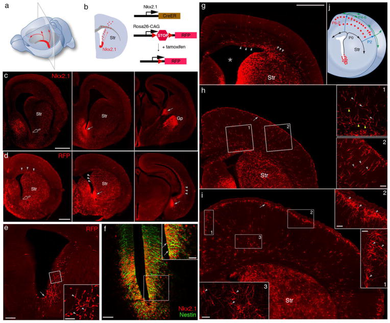
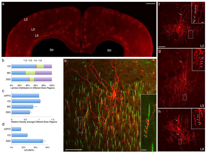
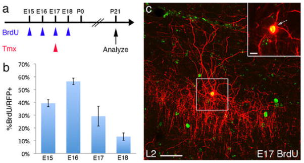
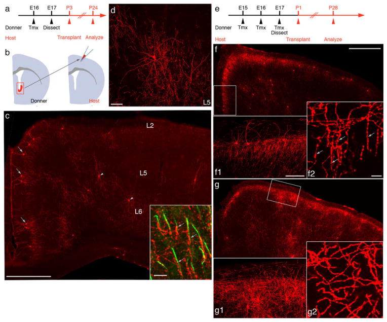
Diverse γ-aminobutyric acid-releasing interneurons regulate the functional organization of cortical circuits and derive from multiple embryonic sources. It remains unclear to what extent embryonic origin influences interneuron specification and cortical integration because of difficulties in tracking defined cell types. Here, we followed the developmental trajectory of chandelier cells (ChCs), the most distinct interneurons that innervate the axon initial segment of pyramidal neurons and control action potential initiation. ChCs mainly derive from the ventral germinal zone of the lateral ventricle during late gestation and require the homeodomain protein Nkx2.1 for their specification. They migrate with stereotyped routes and schedule and achieve specific laminar distribution in the cortex. The developmental specification of this bona fide interneuron type likely contributes to the assembly of a cortical circuit motif.


Figures

Fig. 1
Fig. 1
Nkx2.1+ progenitors in the embryonic VGZ are a novel source of cortical interneurons. (A) NKX2.1 expression (red) in middle-caudal regions of the late embryonic VGZ. Arrows indicate the migration of VGZ-derived cells toward the dorsal and rostral cortex. (B) A coronal section from a region indicated in (A). Red dots represent VGZ-derived cells migrating toward the cortex. Genetic fate mapping strategy is depicted to the right. In an Nkx2.1CreER;Ai9 mouse, Cre-mediated recombination is induced by tamoxifen and activates RFP expression. Str, striatum. (C) Immunofluorescence showing NKX2.1 protein (arrows) in the middle-to-caudal region (middle and right panels) but not rostral region (open arrow in left panel) of E17 VGZ. (D) Nkx2.1CreER;Ai9 mice induced at E17 and examined at E18. RFP-labeled progenitors were abundant in the middle-caudal regions (solid arrows) but not rostral region (open arrow) of VGZ. RFP+ migrating cells (arrowheads) were found not only in midcaudal sections but also in the rostral section. (E) Coronal section of an E18 brain induced by low-dose tamoxifen. More sparse VGZ progenitors (arrow) and postmitotic cells (arrowheads in inset) are labeled, giving a clearer view of the migratory stream along the lateral wall of the ventricle. (F) Colocalization (arrow) of NKX2.1 and Nestin in VGZ progenitors shown by double immunostaining. (G) Coronal section of a P0 brain induced at E17. Migrating cells emerge from the dorso-lateral wall of the ventricle (star) and split into lateral and medial stream (arrowheads) within the cortical subventricular zone. (H) Coronal section of a P2 brain induced at E17. Migrating cells, each with a characteristic leading process (arrowheads in insets) pass through cortical plate into L1 (arrow). (Insets) Tangentially migrating cells in the subventricular and intermediate zone (yellow arrowheads, H1) and radially migrating cells in the cortical plate (light gray arrowheads, H2). (I) Coronal section of a P7 brain induced at E17. RFP+ cells descend from L1 into the cortex and settle at the L1/L2 border (arrow), forming dense clusters, and start to differentiate (arrowheads in I1 and I2). Deep-layer RFP+ cells (arrowheads in I3) also settle and show sign of differentiation by this stage but their migration route is unclear. (J) Diagram depicting the migration route and schedule of VGZ-derived cortical cells. Scale bars: 500 μm in (C), (D), and (G to I); 200 μm in (E); 50 μm in (F) and insets of (E), (H), and (I); and 25 μm in inset of (F).
Fig. 2
Fig. 2
Areal and laminar distribution of ChCs in neocortex. (A) Coronal section showing the overall distribution of RFP+ neurons at P28 after E17 induction of an Nkx2.1CreER;Ai9 mouse. (B) Laminar distribution of RFP+ cells in cingulate (CC), motor (MC) and somatosensory (SSC) cortices. (C) L2 RFP+ cells are more abundant in medial frontal cortices (mPFC, CC) compared with sensory cortices (SSC); ~70% and 90% of RFP+ cells are ChCs in SSC and mPFC, respectively. (D) C-expression of PV in L2 RFP+ cells varies greatly across cortical areas. (E) Colabeling of pyramidal cell AIS [phosphorylated inhibitor of nuclear factor κB (IκB), green] and an L2 ChC. Terminal cartridges align with AISs and target the distal portion of AISs (arrowheads in inset). (F) An L2 ChC with characteristic chandelier-like axon arbor and terminal cartridges bearing strings of synaptic boutons (arrowheads in inset). Note the prominent L1 dendrites (arrow). (G) An L5 ChC with characteristic chandelier-like axon arbor and terminal cartridges (arrowheads in inset). Note the relatively straight apical dendrites (arrow). (H) An L6 ChC with dense local axon arbor and terminal cartridges (arrowheads in inset); its dendrites (arrows) extend above and below the soma. Scale bars: 500 μm in (A), 50 μm in (E) and (F–H), and 5 μm in insets of (E) and (F–H).
Fig. 3
Fig. 3
Temporal profile of ChC generation in the NKX2.1 lineage. (A) Scheme of ChC birth dating. In an Nkx2.1CreER;Ai9 mouse, NKX2.1 lineage cells are labeled by tamoxifen (Tmx) induction at E17 (red arrowhead), paired with a single BrdU injection at the indicated date (blue arrowheads). Mice were analyzed for colocalization after P21. (B) The temporal profile of L2 ChC generation in the cingulate cortex, with a peak at E16. (C) A BrdU-labeled L2 ChC. (D and E) L5 and L6 ChCs are also born in the late embryonic stage. A ChC in L5 (D) and L6 (E) labeled by BrdU at E17, tamoxifen-induced at E17, and analyzed at P28. Insets are single confocal sections. Scale bars: 50 μm in (C) to (E) and 10 μm in insets.
Fig. 4
Fig. 4
NKX2.1 in VGZ progenitors is necessary for the specification of ChCs. (A) Scheme of transplantation experiment. Nkx2.1CreER;Ai9 donors are induced at E16 with tamoxifen (Tmx); RFP-labeled cells from VGZ are dissected at E17 and transplanted to the somatosensory cortex of P3 wild-type host pups and analyzed at P24. (B) The transplantation of RFP+ VGZ cells to a wild-type brain. (C) Coronal section of a P24 host cortex in which ChCs differentiated from transplanted RFP+ VGZ cells. L2 (arrows) and L5 ChCs (arrowheads) are readily identified. The laminar distribution of transplanted ChCs closely resembles that of endogenous ChCs, and L2 ChCs extend characteristic L1 dendrites and local axon arbors. Inset, transplanted ChC axon terminals (arrows) innervate PyNs at AIS (phosphorylated IκB, green) (D) High-magnification view of a transplanted L5 ChC with characteristic dendritic and axon morphology. (E) Scheme of transplantation from Nkx2.1CreER/fix(Nkx2.1KO);Ai9 embryos. Animals are induced three times from E15 to E17, and RFP+ VGZ cells are transplanted into P1 wild-type pups. In the same set of experiments, Nkx2.1CreER/+(Nkx2.1+/−);Ai9 mice were used as controls. (F) Coronal section of a P28 host brain transplanted with Nkx2.1+/− VGZ cells. ChCs accumulate in L2-L1 border, as well as in L5. Higher-magnification view of a boxed region shows dense vertical-oriented axon terminals (F1), with characteristic strings of boutons (F2). (G) Neurons derived from Nkx2.1KO VGZ cells accumulate in L2 and L5, but fail to differentiate into ChCs. Higher-magnification view of the boxed region shows a lack of L1 dendrites and vertical oriented axon terminals (G1), with almost a complete absence of terminal cartridges, (G2). Scale bars: 500 μm in (C), (F), (G); 100 μm in (F1) and (G1); 10 μm in (F2) and (G2).

Comment in

References

    1. Markram H, et al. Interneurons of the neocortical inhibitory system. Nat Rev Neurosci. 2004;5:793. doi: 10.1038/nrn1519. - DOI - PubMed
    1. Gelman DM, Marín O. Generation of interneuron diversity in the mouse cerebral cortex. Eur J Neurosci. 2010;31:2136. doi: 10.1111/j.1460-9568.2010.07267.x. - DOI - PubMed
    1. Ascoli GA, et al. Petilla terminology: Nomenclature of features of GABAergic interneurons of the cerebral cortex. Nat Rev Neurosci. 2008;9:557. doi: 10.1038/nrn2402. - DOI - PMC - PubMed
    1. Szentágothai J, Arbib MA. Conceptual models of neural organization. Neurosci Res Program Bull. 1974;12:305. - PubMed
    1. Somogyi P. A specific ‘axo-axonal’ interneuron in the visual cortex of the rat. Brain Res. 1977;136:345. doi: 10.1016/0006-8993(77)90808-3. - DOI - PubMed

Publication types

MeSH terms