. 2012 Dec 21;338(6114):1631-4.
doi: 10.1126/science.1227270. Epub 2012 Nov 22.

Organization of the influenza virus replication machinery

Affiliations

Organization of the influenza virus replication machinery

Arne Moeller et al. Science. .

Abstract

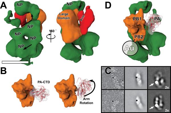
Influenza virus ribonucleoprotein complexes (RNPs) are central to the viral life cycle and in adaptation to new host species. RNPs are composed of the viral genome, viral polymerase, and many copies of the viral nucleoprotein. In vitro cell expression of all RNP protein components with four of the eight influenza virus gene segments enabled structural determination of native influenza virus RNPs by means of cryogenic electron microscopy (cryo-EM). The cryo-EM structure reveals the architecture and organization of the native RNP, defining the attributes of its largely helical structure and how polymerase interacts with nucleoprotein and the viral genome. Observations of branched-RNP structures in negative-stain electron microscopy and their putative identification as replication intermediates suggest a mechanism for viral replication by a second polymerase on the RNP template.


Figures

Fig. 1
Fig. 1. Cryo-EM reconstruction of the influenza virus ribonucleoprotein complex
(A) Composite model of cryo-EM reconstructions of the three regions of the RNP. (B) Cartoon representation of the RNP organization. The large domain of polymerase is shown in orange with the arm domain in red. Nucleoproteins are shown in green and RNA in blue. (C) Reconstruction of the central filament region using helical symmetry. (D) Single protomers from the NP crystal structure () were fitted into the EM density. The arrangement of the NP (light-blue for descending strand and dark blue for ascending strand) within the filament creates a periodic box type arrangement formed from four NPs with a region of low density or dimple in the center of the box. This box-like feature is also easily identifiable in our reconstructions of the loop and polymerase end regions. Arrows indicate RNA polarity. Scale bar represents 10 nm.
Fig. 2
Fig. 2. The influenza virus RNA polymerase and its interactions at the RNP terminus
(A) The RNP-bound polymerase large domain (orange) caps the central RNP filament region. A single NP from the second strand that has emerged from the central filament follows the 3′ RNA strand as it loops behind polymerase to contact the PA CTD (red). (B) Cryo-EM reconstruction of the free polymerase (left) shows it consists of a large domain (orange) and an arm containing the PA CTD (red) (). The arm domain conformation in the RNP is accommodated by a rotation about a pivot near the base of the arm (right). (C) 2-D averages and raw images of negatively stained RNPs labeled with 5 nm Nanogold (upper and middle panels respectively) localize the PB2 C terminus to the bottom of the large domain near the NP contact site. The lower panels show 2-D projections of 21 Å RNP-bound polymerase with an additional circle (indicated with an arrow) below the polymerase-large domain corresponding to the size of the 5 nm Nanogold for comparison with labeled 2-D class averages. (D) The composite image of the RNP polymerase end has been labeled to indicate putative subunit locations based on the PA CTD docking, Nanogold labeling of the PB2 C-terminus and structural homology with reovirus polymerase. The location of the 5 nm Nanogold labeling the PB2 C-terminus is shown as a circle labeled Au. Because the reconstruction is centered on the polymerase and the RNP is flexible, additional density corresponding to distal regions of poorly aligned RNP filaments is visible beneath the well-ordered components.
Fig. 3
Fig. 3. Models for influenza virus RNA synthesis
(A) In the resting RNP, polymerase is bound to both 5′ and 3′ termini, as would be expected in virions. The polymerase large domain and arm domain are colored orange and red respectively. The nucleoprotein is in green and the genomic RNA is in light blue for 3′ end and dark blue for the 5′ end. The active site (*) and RNA polarity were identified using structural homology with the reovirus λ3 polymerase (fig S13). (B) Viral transcription of mRNA is carried out by the resident polymerase acting. In this process, template RNA is pulled up from beneath polymerase, passed through the active site where it is transcribed into capped mRNA (black) and then re-encapsidated into a NP-RNA complex, which coils up to form an RNP-like structure. (C) Replication of viral RNA is carried out by a second polymerase leading to nascent RNP formation. The second replicating polymerase binds a newly synthesized 5′ end and initiates encapsidation of the viral genome by NP in a 5′ to 3′ manner leading to nascent RNP formation.
Fig. 4
Fig. 4. Formation of nascent RNP during replication. (Upper panel)
Electron micrographs of the negatively stained RNP sample at high dilution reveal branched RNPs predicted to be replication intermediates. (Lower panel) Interpretation of the branched RNPs in the upper panel showing how replication of the viral RNA in trans by a second polymerase (red) could result in nascent RNP complexes (dark green) branching from the template RNP (light green). As the nascent RNP moves away from and then back towards the template RNP polymerase, that is, 3′ to 5′ along the genomic template, it would elongate the complementary RNA and extends its length. Scale bar corresponds to 100 nm.

Comment in

References

    1. Fields BN, Knipe DM, Howley PM. Fields' Virology. Wolters Kluwer Health/Lippincott Williams & Wilkins; 2007.
    1. Neumann G, Noda T, Kawaoka Y. Emergence and pandemic potential of swine-origin H1N1 influenza virus. Nature. 2009;459:931–939. - PMC - PubMed
    1. Hatta M, Gao P, Halfmann P, Kawaoka Y. Molecular basis for high virulence of Hong Kong H5N1 influenza A viruses. Science. 2001;293:1840–1842. - PubMed
    1. Gabriel G, et al. The viral polymerase mediates adaptation of an avian influenza virus to a mammalian host. Proc. Natl. Acad. Sci. USA. 2005;102:18590–18595. - PMC - PubMed
    1. Compans RW, Content J, Duesberg PH. Structure of the ribonucleoprotein of influenza virus. J. Virol. 1972;10:795–800. - PMC - PubMed

Publication types

MeSH terms

Associated data