

Phosphatidylinositol 3-kinase-γ signaling promotes Campylobacter jejuni-induced colitis through neutrophil recruitment in mice
- PMID: 23180818
- PMCID: PMC3529774
- DOI: 10.4049/jimmunol.1201825
Phosphatidylinositol 3-kinase-γ signaling promotes Campylobacter jejuni-induced colitis through neutrophil recruitment in mice
Abstract
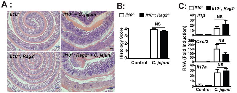
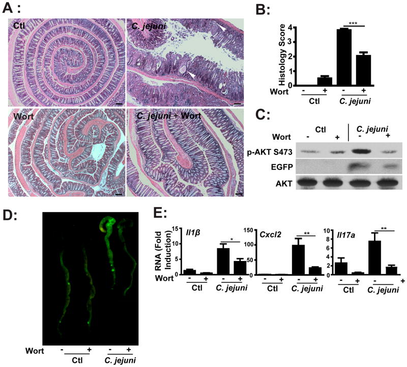
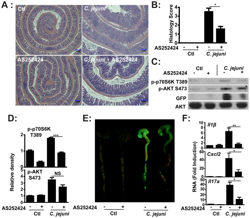
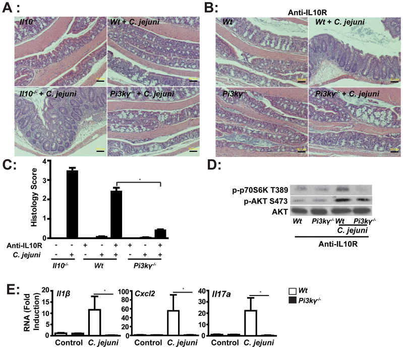
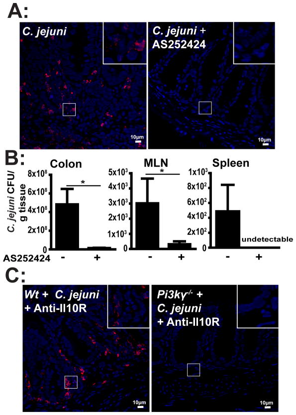
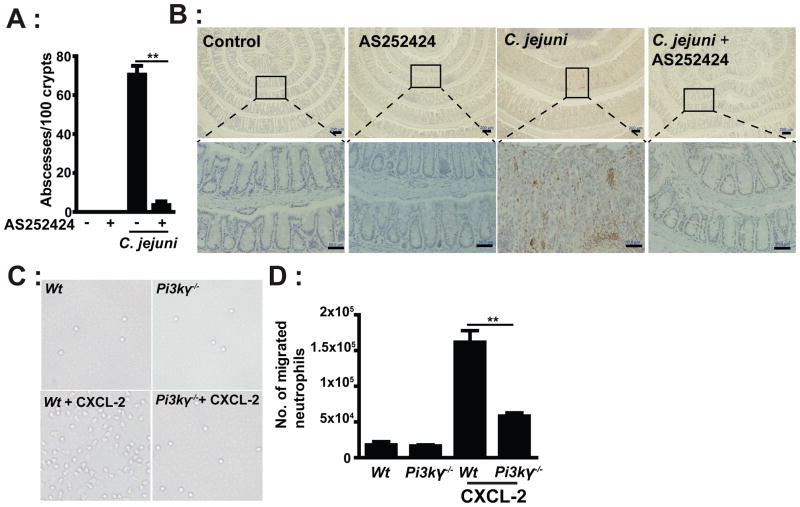
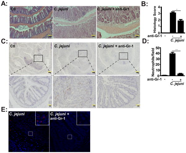
Crypt abscesses caused by excessive neutrophil accumulation are prominent features of human campylobacteriosis and its associated pathology. The molecular and cellular events responsible for this pathological situation are currently unknown. We investigated the contribution of PI3K-γ signaling in Campylobacter jejuni-induced neutrophil accumulation and intestinal inflammation. Germ-free and specific pathogen-free Il10(-/-) and germ-free Il10(-/-);Rag2(-/-) mice were infected with C. jejuni (10(9) CFU/mouse). PI3K-γ signaling was manipulated using either the pharmacological PI3K-γ inhibitor AS252424 (i.p. 10 mg/kg daily) or genetically using Pi3k-γ(-/-) mice. After up to 14 d, inflammation was assessed histologically and by measuring levels of colonic Il1β, Cxcl2, and Il17a mRNA. Neutrophils were depleted using anti-Gr1 Ab (i.p. 0.5 mg/mouse/every 3 d). Using germ-free Il10(-/-);Rag2(-/-) mice, we observed that innate immune cells are the main cellular compartment responsible for campylobacteriosis. Pharmacological blockade of PI3K-γ signaling diminished C. jejuni-induced intestinal inflammation, neutrophil accumulation, and NF-κB activity, which correlated with reduced Il1β (77%), Cxcl2 (73%), and Il17a (72%) mRNA accumulation. Moreover, Pi3k-γ(-/-) mice pretreated with anti-IL-10R were resistant to C. jejuni-induced intestinal inflammation compared with Wt mice. This improvement was accompanied by a reduction of C. jejuni translocation into the colon and extraintestinal tissues and by attenuation of neutrophil migratory capacity. Furthermore, neutrophil depletion attenuated C. jejuni-induced crypt abscesses and intestinal inflammation. Our findings indicate that C. jejuni-induced PI3K-γ signaling mediates neutrophil recruitment and intestinal inflammation in Il10(-/-) mice. Selective pharmacological inhibition of PI3K-γ may represent a novel means to alleviate severe cases of campylobacteriosis, especially in antibiotic-resistant strains.
Conflict of interest statement
Figures
Similar articles
-
Nucleotide-binding oligomerization domain-containing protein 2 controls host response to Campylobacter jejuni in Il10-/- mice.J Infect Dis. 2014 Oct 1;210(7):1145-54. doi: 10.1093/infdis/jiu148. Epub 2014 Mar 11. J Infect Dis. 2014. PMID: 24620022 Free PMC article.
-
Campylobacter jejuni induces colitis through activation of mammalian target of rapamycin signaling.Gastroenterology. 2012 Jan;142(1):86-95.e5. doi: 10.1053/j.gastro.2011.09.042. Epub 2011 Oct 1. Gastroenterology. 2012. PMID: 21963787 Free PMC article.
-
Microbiota attenuates chicken transmission-exacerbated campylobacteriosis in Il10-/- mice.Sci Rep. 2020 Nov 30;10(1):20841. doi: 10.1038/s41598-020-77789-2. Sci Rep. 2020. PMID: 33257743 Free PMC article.
-
IL-23 Contributes to Campylobacter jejuni-Induced Intestinal Pathology via Promoting IL-17 and IFNγ Responses by Innate Lymphoid Cells.Front Immunol. 2021 Jan 6;11:579615. doi: 10.3389/fimmu.2020.579615. eCollection 2020. Front Immunol. 2021. PMID: 33488580 Free PMC article.
-
Intestinal microbiota shifts towards elevated commensal Escherichia coli loads abrogate colonization resistance against Campylobacter jejuni in mice.PLoS One. 2012;7(5):e35988. doi: 10.1371/journal.pone.0035988. Epub 2012 May 1. PLoS One. 2012. PMID: 22563475 Free PMC article.
Cited by
-
Protein kinases are potential targets to treat inflammatory bowel disease.World J Gastrointest Pharmacol Ther. 2014 Nov 6;5(4):209-17. doi: 10.4292/wjgpt.v5.i4.209. World J Gastrointest Pharmacol Ther. 2014. PMID: 25374761 Free PMC article. Review.
-
Nucleotide-binding oligomerization domain-containing protein 2 controls host response to Campylobacter jejuni in Il10-/- mice.J Infect Dis. 2014 Oct 1;210(7):1145-54. doi: 10.1093/infdis/jiu148. Epub 2014 Mar 11. J Infect Dis. 2014. PMID: 24620022 Free PMC article.
-
Contrasting immune responses mediate Campylobacter jejuni-induced colitis and autoimmunity.Mucosal Immunol. 2014 Jul;7(4):802-17. doi: 10.1038/mi.2013.97. Epub 2013 Nov 13. Mucosal Immunol. 2014. PMID: 24220299 Free PMC article.
-
The fibronectin-binding motif within FlpA facilitates Campylobacter jejuni adherence to host cell and activation of host cell signaling.Emerg Microbes Infect. 2013 Oct;2(10):e65. doi: 10.1038/emi.2013.65. Epub 2013 Oct 9. Emerg Microbes Infect. 2013. PMID: 26038437 Free PMC article.
-
Tanshinone IIA Protects against Dextran Sulfate Sodium- (DSS-) Induced Colitis in Mice by Modulation of Neutrophil Infiltration and Activation.Oxid Med Cell Longev. 2016;2016:7916763. doi: 10.1155/2016/7916763. Epub 2016 Jan 3. Oxid Med Cell Longev. 2016. PMID: 26881040 Free PMC article.
References
-
- CDC. Incidence of laboratory-confirmed bacterial and parasitic infections, and postdiarrheal hemolytic uremic syndrome (HUS), by year and pathogen, Foodborne Diseases Active Surveillance Network (FoodNet), United States, 1996 – 2011. Centers for Disease Control and Prevention; 2012. http://www.cdc.gov/foodnet/data/trends/tables/table2a-b.html#table-2b.
-
- Blaser MJ. Epidemiologic and clinical features of Campylobacter jejuni infections. J Infect Dis. 1997;176(Suppl 2):S103–105. - PubMed
-
- Nachamkin I. Chronic effects of Campylobacter infection. Microbes Infect. 2002;4:399–403. - PubMed
-
- Mortensen NP, Kuijf ML, Ang CW, Schiellerup P, Krogfelt KA, Jacobs BC, van Belkum A, Endtz HP, Bergman MP. Sialylation of Campylobacter jejuni lipo-oligosaccharides is associated with severe gastro-enteritis and reactive arthritis. Microbes Infect. 2009;11:988–994. - PubMed
-
- Gradel KO, Nielsen HL, Schønheyder HC, Ejlertsen T, Kristensen B, Nielsen H. Increased Short- and Long-Term Risk of Inflammatory Bowel Disease After Salmonella or Campylobacter Gastroenteritis. Gastroenterology. 2009;137:495–501. - PubMed
Publication types
MeSH terms
Substances
Grants and funding
LinkOut - more resources
Full Text Sources
Molecular Biology Databases
