. 2013 Mar;27(3):1001-11.
doi: 10.1096/fj.12-219618. Epub 2012 Nov 24.

Spns2, a transporter of phosphorylated sphingoid bases, regulates their blood and lymph levels, and the lymphatic network

Affiliations

Spns2, a transporter of phosphorylated sphingoid bases, regulates their blood and lymph levels, and the lymphatic network

Masayuki Nagahashi et al. FASEB J. 2013 Mar.

Abstract

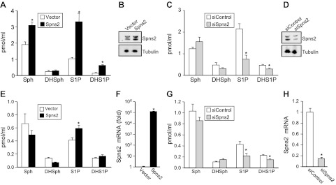
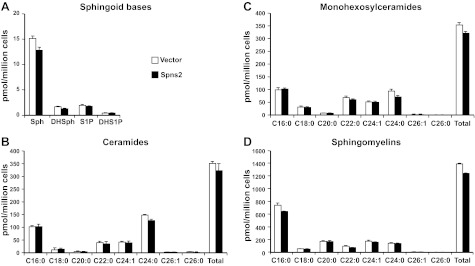
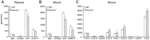
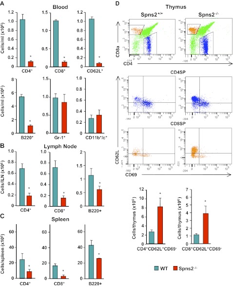
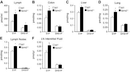
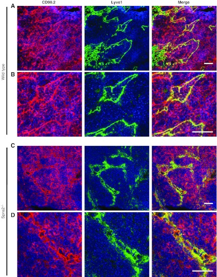
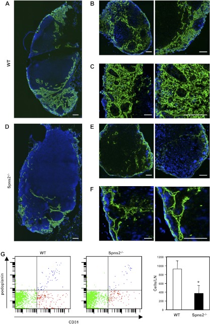
Sphingosine-1-phosphate (S1P), a ligand for 5 specific receptors, is a potent lipid mediator that plays important roles in lymphocyte trafficking and immune responses. S1P is produced inside cells and therefore must be secreted to exert its effects through these receptors. Spinster 2 (Spns2) is one of the cell surface transporters thought to secrete S1P. We have shown that Spns2 can export endogenous S1P from cells and also dihydro-S1P, which is active at all cell surface S1P receptors. Moreover, Spns2 mice have decreased levels of both of these phosphorylated sphingoid bases in blood, accompanied by increases in very long chain ceramide species, and have defective lymphocyte trafficking. Surprisingly, levels of S1P and dihydro-S1P were increased in lymph from Spns2 mice as well as in specific tissues, including lymph nodes, and interstitial fluid. Moreover, lymph nodes from Spns2 mice have aberrant lymphatic sinus that appeared collapsed, with reduced numbers of lymphocytes. Our data suggest that Spns2 is an S1P transporter in vivo that plays a role in regulation not only of blood S1P but also lymph node and lymph S1P levels and consequently influences lymphocyte trafficking and lymphatic vessel network organization.


Figures

Figure 1.
Figure 1.
Spns2 transports S1P and dihydro-S1P from cells. A–D) Spns2 was overexpressed (A, B) or down-regulated (C, D) in HEK 293 cells. Then 48 h later, cells were washed, and culture medium was replaced with medium containing a phosphatase inhibitor. After 2 h, medium was collected and sphingosine (Sph), dihydrosphingosine (DHSph), S1P, and dihydro-S1P (DHS1P) were measured by LC-ESI-MS/MS (A, C). Data are means ± sd of triplicate determinations. *P < 0.05. B, D) In duplicate cultures, Spns2 expression was determined with anti-Spns2 antibody (Sigma-Aldrich). Blots were stripped and reprobed with anti-tubulin antibody to ensure equal loading and transfer. E–H) Similar results were found in 2 additional experiments. Spns2 was overexpressed (E, F) or down-regulated (G, H) in TIME cells. Sphingolipids were measured by LC-ESI-MS/MS (E, G), and mRNA levels of Spns2 were determined by quantitative real-time PCR and normalized to glyceraldehyde-3-phosphate dehydrogenase (F, H).
Figure 2.
Figure 2.
Overexpression of Spns2 does not alter cellular levels of S1P and dihydro-S1P or other sphingolipids. HEK 293 cells transfected with control or Spns2 were washed, and culture medium was replaced with medium containing phosphatase inhibitor. After 2 h, lipids were extracted from cells, and dihydrosphingosine, dihydro-S1P, sphingosine, and S1P (A), different chain length species of ceramide (B), monohexosylceramide (C), and sphingomyelin (D) were determined by LC-ESI-MS/MS. Numbers indicate chain length, followed by the number of double bonds in the fatty acid. Data are averages of triplicate determinations and are expressed as picomoles of lipid per million cells. Similar results were found in two additional experiments.
Figure 3.
Figure 3.
Blood levels of S1P and dihydro-S1P are reduced, whereas very long chain ceramides are increased in Spns2-knockout mice. Lipids were extracted from plasma (A) and whole blood (B, C) from wild-type (WT) and Spns2-knockout mice and dihydrosphingosine, dihydro-S1P, sphingosine, and S1P (A, B), and ceramide species (C) were determined by LC-ESI-MS/MS. Numbers indicate chain length followed by the number of double bonds in the fatty acid. *P < 0.05.
Figure 4.
Figure 4.
Aberrant lymphocyte trafficking in Spns2−/− mice. Lymphocytes of wild-type (WT) and Spns2−/− mice were analyzed by flow cytometry. A) Number of circulating CD3+ T cells that are CD4+, CD8+, or CD62L+; B220+ B cells; Gr-1+ granulocytes; and CD11b+/c+ dendritic cells. B, C) Total number of CD3+ T cells that are CD4+, CD8+, or CD62L+ and B220+ B cells in lymph nodes (B) and spleen (C). D) Flow cytometry analysis of wild-type and Spns2−/− thymocytes. Single-positive CD4+ and CD8+ T cells were gated for the maturation markers CD69 and CD62L. Representative flow cytometry plots (top panels) and total number of CD4+CD62L+CD69 and CD4+CD62L+CD69 T cells (bottom panels) are shown. *P < 0.01.
Figure 5.
Figure 5.
Increased levels of S1P in lymph and tissues of Spns2−/− mice. Lipids were extracted from lymph (A), colon (B), liver (C), lung (D), lymph nodes (E), and lymph node (LN) interstitial fluid (F) from wild-type (WT) and Spns2-knockout mice, and S1P and dihydro-S1P (DHS1P) were determined by LC-ESI-MS/MS. *P < 0.05.
Figure 6.
Figure 6.
Impaired T-cell egress from lymph nodes in Spns2-deficient mice. Lymph nodes from wild-type (A, B) and Spns2−/− mice (C, D) were immunostained to visualize LECs with LYVE-1 (green), T cells with CD90.2 (red), and nuclei with Hoechst (blue). Confocal fluorescent images at ×40 (A, C) and ×95 (B, D). Scale bars = 100 μm.
Figure 7.
Figure 7.
Lack of Spns2 disrupts the lymphatic vessel network in lymph nodes. A–F) Immunofluorescent analysis of lymph nodes from wild-type (WT) littermates and Spns2−/− mice stained for LYVE-1 (green) and Hoechst (blue). Confocal images of axillary lymph nodes from wild-type (A–C) and Spns−/− mice (D–F), showing lymphatic sinus stained with antibody against LYVE-1 with low magnification using tile scan technology (A, B, D, E) and with high magnification (C, F). Scale bars = 100 μm. G) Single-cell suspensions were prepared from axillary lymph nodes of wild-type and Spns2−/− mice by digestion with collagenase, and total numbers of LECs were quantified by FACS analysis. Data are means ± se of triplicate determinations.

Similar articles

Cited by

References

    1. Spiegel S., Milstien S. (2011) The outs and the ins of sphingosine-1-phosphate in immunity. Nat. Rev. Immunol. 11, 403–415 - PMC - PubMed
    1. Cyster J. G., Schwab S. R. (2012) Sphingosine-1-phosphate and lymphocyte egress from lymphoid organs. Annu. Rev. Immunol. 30, 69–94 - PubMed
    1. Mitra P., Oskeritzian C. A., Payne S. G., Beaven M. A., Milstien S., Spiegel S. (2006) Role of ABCC1 in export of sphingosine-1-phosphate from mast cells. Proc. Natl. Acad. Sci. U. S. A. 103, 16394–16399 - PMC - PubMed
    1. Sato K., Malchinkhuu E., Horiuchi Y., Mogi C., Tomura H., Tosaka M., Yoshimoto Y., Kuwabara A., Okajima F. (2007) Critical role of ABCA1 transporter in sphingosine 1-phosphate release from astrocytes. J. Neurochem. 103, 2610–2619 - PubMed
    1. Takabe K., Kim R. H., Allegood J. C., Mitra P., Ramachandran S., Nagahashi M., Harikumar K. B., Hait N. C., Milstien S., Spiegel S. (2010) Estradiol induces export of sphingosine 1-phosphate from breast cancer cells via ABCC1 and ABCG2. J. Biol. Chem. 285, 10477–10486 - PMC - PubMed

Publication types