. 2012 Nov 26:9:257.
doi: 10.1186/1742-2094-9-257.

670-nm light treatment reduces complement propagation following retinal degeneration

Affiliations

670-nm light treatment reduces complement propagation following retinal degeneration

Matt Rutar et al. J Neuroinflammation. .

Abstract

Aim: Complement activation is associated with the pathogenesis of age-related macular degeneration (AMD). We aimed to investigate whether 670-nm light treatment reduces the propagation of complement in a light-induced model of atrophic AMD.

Methods: Sprague-Dawley (SD) rats were pretreated with 9 J/cm(2) 670-nm light for 3 minutes daily over 5 days; other animals were sham treated. Animals were exposed to white light (1,000 lux) for 24 h, after which animals were kept in dim light (5 lux) for 7 days. Expression of complement genes was assessed by quantitative polymerase chain reaction (qPCR), and immunohistochemistry. Counts were made of C3-expressing monocytes/microglia using in situ hybridization. Photoreceptor death was also assessed using outer nuclear layer (ONL) thickness measurements, and oxidative stress using immunohistochemistry for 4-hydroxynonenal (4-HNE).

Results: Following light damage, retinas pretreated with 670-nm light had reduced immunoreactivity for the oxidative damage maker 4-HNE in the ONL and outer segments, compared to controls. In conjunction, there was significant reduction in retinal expression of complement genes C1s, C2, C3, C4b, C3aR1, and C5r1 following 670 nm treatment. In situ hybridization, coupled with immunoreactivity for the marker ionized calcium binding adaptor molecule 1 (IBA1), revealed that C3 is expressed by infiltrating microglia/monocytes in subretinal space following light damage, which were significantly reduced in number after 670 nm treatment. Additionally, immunohistochemistry for C3 revealed a decrease in C3 deposition in the ONL following 670 nm treatment.

Conclusions: Our data indicate that 670-nm light pretreatment reduces lipid peroxidation and complement propagation in the degenerating retina. These findings have relevance to the cellular events of complement activation underling the pathogenesis of AMD, and highlight the potential of 670-nm light as a non-invasive anti-inflammatory therapy.


Figures

Figure 1
Figure 1
Measurements of outer nuclear layer (ONL) thickness following 670-nm light treatment and bright continuous white light (BCL) exposure. The thickness of the ONL decreased significantly following exposure to BCL, compared to both dim-reared and dim-reared + 670 nm groups (P <0.05). In comparison, the ONL was significantly thicker in animals treated with 670-nm light prior to BCL exposure (P <0.05). Dim reared n = 3, dim reared + 670 nm n = 3, light damage n = 3, light damage + 670-nm light n = 3; error bars represent SEM. *Significant change using analysis of variance (ANOVA) with Tukey’s post test where P <0.05.
Figure 2
Figure 2
Immunoreactivity (IR) for 4-hydroxynonenal (4-HNE; red) in the retina following 670-nm light treatment and exposure to bright continuous white light (BCL). (A,B) IR for 4-HNE was faintly detected in the inner (IS) and outer segments (OS) of both untreated (A) and 670-nm light-treated (B) dim-reared animals (arrows). (C,D) IR for 4-HNE was more intense in the IS and OS (arrow) following exposure to BCL. (E) Treatment with 670-nm light prior to BCL resulted in a marked reduction in IR for 4-HNE (arrow), compared to light damage alone (C). (F) Negative controls showed no specific staining for 4-HNE following BCL exposure. ONL = outer nuclear layer; OS = outer segments.
Figure 3
Figure 3
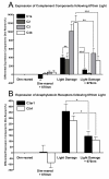
Expression of complement-related genes in the retina by quantitative polymerase chain reaction (qPCR) following 670-nm light treatment and bright continuous white light (BCL) exposure. (A,B) The expression of complement components C1s, C2, C3, C4b (A) and receptors C3aR1, C5r1 (B) increased significantly following BCL relative to dim-reared animals (P <0.05). In animals treated with 670-nm light prior to BCL exposure, the expression of all complement-related genes assessed was substantially reduced, compared to those exposed to BCL alone (P <0.05). Dim-reared animals exposed 670-nm light showed no significant modulation of complement gene expression relative to controls (P >0.05). Dim reared n = 3, dim reared + 670 nm n = 3, light damage n = 3, light damage + 670-nm light n = 3; error bars represent SEM. *Significant change using analysis of variance (ANOVA) with Tukey’s post test where P <0.05.
Figure 4
Figure 4
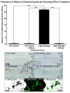
In situ hybridization for C3 mRNA in the retina following 670-nm light and bright continuous white light (BCL) exposure. (A-E) Representative images from the superior mid-periphery show in situ hybridization for C3 mRNA in the retina. Retinas from dim-reared (A) and 670-nm light-treated dim-reared (B) animals showed no staining for C3 mRNA in the retina except for infrequent C3-expressing nuclei associated with the retina vasculature (data not shown). In BCL-exposed animals (C-D) C3-expressing nuclei were more numerous in the outer nuclear layer (ONL) and outer segments within the lesion area, while none were observed in those treated with 670-nm light prior to BCL exposure (E). (F,G) C3 expression (dark grey) in sections counterimmunolabelled with anti-IBA1 (green), showing immunoreactivity in C3-expressing nuclei within the degenerating ONL (arrows) following BCL. Histogram: Quantification of C3-expressing nuclei per retina showed a dramatic increase from near zero in dim-reared animals to 26.7 following BCL exposure (P <0.05). In contrast, the number of C3-expressing nuclei was reduced to near zero in animals pretreated with 670-nm light following BCL (P <0.05). Dim reared n = 3, dim reared + 670-nm light n = 3, light damage n = 3, light damage + 670-nm light n = 3; error bars represent SEM. *Significant change using analysis of variance (ANOVA) with Tukey’s post test where P <0.05.
Figure 5
Figure 5
Immunoreactivity (IR) for C3 (red) in the retina following 670-nm light treatment and exposure to bright continuous white light (BCL). (A-F) Representative images of C3 immunoreactivity taken from the superior mid-periphery. (A,B) IR for C3 was faintly detected in retinal vasculature (arrows) of dim-reared retinas (A), as well as those treated with 670-nm light (B), at comparable levels of IR. (C-F) Following BCL exposure, IR for C3 was observed in multiple deposits throughout the outer nuclear layer (ONL) in the lesion area (C-D, arrows) and the layer of outer segments at the edge of the lesion (E-F, arrows). (G) C3-IR was vastly reduced in the ONL and outer segments of animals treated with 670-nm light prior to BCL, and appeared similar to dim-reared controls (arrows). (H) Negative controls showed no specific staining for C3 following BCL exposure. (I-L) C3-IR (red) in the outer segments of sections counterimmunolabelled with anti-4-hydroxynonenal (4-HNE) (green), shows colocalization for aggregations of 4-HNE immunoreactivity (arrows) following BCL. C = choroid; ONL = outer nuclear layer; OS = outer segment layer.

References

    1. Ambati J. Age-related macular degeneration and the other double helix. The Cogan Lecture. Invest Ophthalmol Vis Sci. 2011;52:2165–2169. doi: 10.1167/iovs.11-7329. - DOI - PMC - PubMed
    1. Ambati J, Ambati BK, Yoo SH, Ianchulev S, Adamis AP. Age-related macular degeneration: etiology, pathogenesis, and therapeutic strategies. Surv Ophthalmol. 2003;48:257–293. doi: 10.1016/S0039-6257(03)00030-4. - DOI - PubMed
    1. Grisanti S, Tatar O. The role of vascular endothelial growth factor and other endogenous interplayers in age-related macular degeneration. Prog Retin Eye Res. 2008;27:372–390. doi: 10.1016/j.preteyeres.2008.05.002. - DOI - PubMed
    1. Wong WT, Kam W, Cunningham D, Harrington M, Hammel K, Meyerle CB, Cukras C, Chew EY, Sadda SR, Ferris FL. Treatment of geographic atrophy by the topical administration of OT-551: results of a phase II clinical trial. Invest Ophthalmol Vis Sci. 2010;51:6131–6139. doi: 10.1167/iovs.10-5637. - DOI - PMC - PubMed
    1. Anderson DH, Radeke MJ, Gallo NB, Chapin EA, Johnson PT, Curletti CR, Hancox LS, Hu J, Ebright JN, Malek G, Hauser MA, Rickman CB, Bok D, Hageman GS, Johnson LV. The pivotal role of the complement system in aging and age-related macular degeneration: hypothesis re-visited. Prog Retin Eye Res. 2010;29:95–112. doi: 10.1016/j.preteyeres.2009.11.003. - DOI - PMC - PubMed

Publication types

MeSH terms

LinkOut - more resources