Comparative Study
. 2012 Nov 27:9:46.
doi: 10.1186/1743-8977-9-46.

Critical role of surface chemical modifications induced by length shortening on multi-walled carbon nanotubes-induced toxicity

Affiliations
Comparative Study

Critical role of surface chemical modifications induced by length shortening on multi-walled carbon nanotubes-induced toxicity

Cyrill Bussy et al. Part Fibre Toxicol. .

Abstract

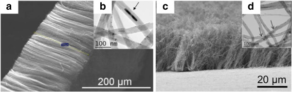
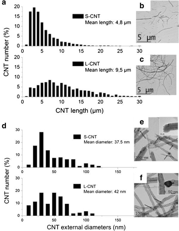
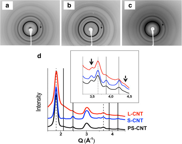
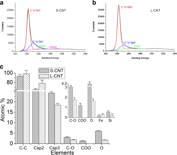
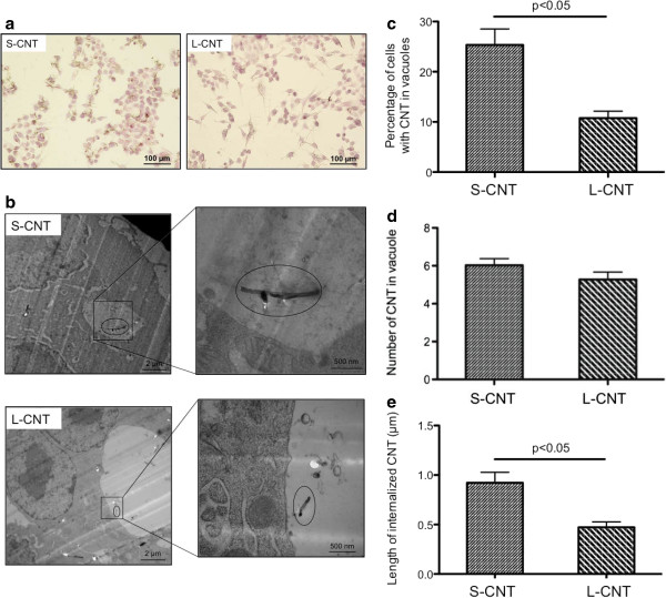
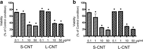
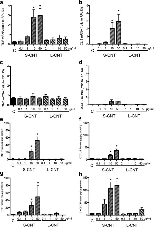
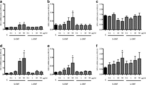
Given the increasing use of carbon nanotubes (CNT) in composite materials and their possible expansion to new areas such as nanomedicine which will both lead to higher human exposure, a better understanding of their potential to cause adverse effects on human health is needed. Like other nanomaterials, the biological reactivity and toxicity of CNT were shown to depend on various physicochemical characteristics, and length has been suggested to play a critical role. We therefore designed a comprehensive study that aimed at comparing the effects on murine macrophages of two samples of multi-walled CNT (MWCNT) specifically synthesized following a similar production process (aerosol-assisted CVD), and used a soft ultrasonic treatment in water to modify the length of one of them. We showed that modification of the length of MWCNT leads, unavoidably, to accompanying structural (i.e. defects) and chemical (i.e. oxidation) modifications that affect both surface and residual catalyst iron nanoparticle content of CNT. The biological response of murine macrophages to the two different MWCNT samples was evaluated in terms of cell viability, pro-inflammatory cytokines secretion and oxidative stress. We showed that structural defects and oxidation both induced by the length reduction process are at least as responsible as the length reduction itself for the enhanced pro-inflammatory and pro-oxidative response observed with short (oxidized) compared to long (pristine) MWCNT. In conclusion, our results stress that surface properties should be considered, alongside the length, as essential parameters in CNT-induced inflammation, especially when dealing with a safe design of CNT, for application in nanomedicine for example.


Figures

Figure 1
Figure 1
Electron microscopy images of PS- and L-CNT. Scanning electron microscopy (SEM, panel a, and c) images and Transmission electron microscopy (TEM, panel b, and d) images of PS-CNT (before length reduction by ultrasonic treatment, panel a and b) and L-CNT (panel c-d). Black arrows point toward iron-based nanoparticles.
Figure 2
Figure 2
Measurement of CNT length and external diameter. Determination of length (panel a) and external diameter (panel d) distributions of S- and L-CNT. TEM images of S-CNT (panel b and e) and L-CNT (panel c and f). Black arrows point toward iron-based nanoparticles.
Figure 3
Figure 3
X-ray diffraction of the different CNT. X-ray diffraction patterns of modified CNT before (panel a, PS-CNT) and after length reduction (panel b, S-CNT), and L-CNT (panel c), powder-like samples being placed in capillaries. The corresponding diffraction diagrams are drawn in (d). The solid and dotted lines indicate positions of diffraction peaks characteristic of iron oxide Fe3O4 nanoparticles and of inter-wall distance in MWCNT, respectively. The solid and dotted arrows in the inset point towards diffraction peaks characteristic of γ and α-iron nanoparticles, while their most intense diffraction peaks are located around 3 Å-1, where CNT contribution is also found. The broad peak below the 002 CNT peak (around 1.83 Å-1) is due to scattering from the glass capillary; its relative intensity with respect to other peaks is meaningless since it only reflects the density of the powder in the capillary.
Figure 4
Figure 4
XPS analysis of S- and L-CNT. XPS spectra C1s core level for (a) S-CNT and (b) L-CNT and (c) corresponding quantitative analyses of the surface chemical composition extracted from the C1s, O1s and Fe2p spectra. Data in (c) are given as mean ± SEM.
Figure 5
Figure 5
Microscopy images of macrophages exposed to S- and L-CNT. Optical microscopy (panel a) and TEM (panel b) images of RAW 264.7 macrophages exposed to 10 μg/mL of S- and L-CNT for 24 hours. Quantification of the percentage of cells with CNT-containing vesicles (panel c). Quantification of the number of CNT inside vesicles (panel d). Quantification of CNT length inside vesicles (panel e). Data are represented as mean ± SEM. P<0.05 between S- and L-CNT.
Figure 6
Figure 6
Viability of macrophages exposed to S- and L-CNT. Quantification of cell viability using WST-1 assay in RAW 264.7 macrophages exposed to 0.1-50 μg/mL of S- or L-CNT for 6 (panel a) or 24 (panel b) hours. *: p<0.05 versus control condition. S-CNT: short CNT. L-CNT: long CNT.
Figure 7
Figure 7
mRNA and protein expression levels of inflammatory cytokines. Quantification of mRNA expression levels of TNF (panel a and c) and CXCL-2 (panel b and d) in RAW 264.7 macrophages exposed to 0.1-50 μg/mL of S- and L-CNT for 6 (panel a and b) or 24 (panel c and d) hours. Quantification of protein expression levels for TNF-α (panel e and g) and CXCL-2 (panel f and h) in RAW 264.7 macrophages exposed to 0.1-50 μg/mL of S- and L-CNT for 6 (panel e and f) or 24 (panel g and h) hours. *: p<0.05 versus control condition. C: Control (unexposed) cells. S-CNT: short CNT. L-CNT: long CNT.
Figure 8
Figure 8
mRNA expression levels of antioxidant systems. Quantification of mRNA expression levels for HO-1 (panel a and d), SOD-2 (panel b and e) and GPX-1 (panel c and f) in RAW 264.7 macrophages exposed to 0.1-50 μg/mL of S- and L-CNT for 6 (panel a-c) or 24 (panel d-f) hours. *: p<0.05 versus control condition. C: Control (unexposed) cells. S-CNT: short CNT. L-CNT: long CNT.

References

    1. Ajayan PM, Tour JM. Materials science: nanotube composites. Nature. 2007;447:1066–1068. doi: 10.1038/4471066a. - DOI - PubMed
    1. Donaldson K, Murphy FA, Duffin R, Poland CA. Asbestos, carbon nanotubes and the pleural mesothelium: a review and the hypothesis regarding the role of long fibre retention in the parietal pleura, inflammation and mesothelioma. Part Fibre Toxicol. 2010;7:5. doi: 10.1186/1743-8977-7-5. - DOI - PMC - PubMed
    1. Helland A, Wick P, Koehler A, Schmid K, Som C. Reviewing the environmental and human health knowledge base of carbon nanotubes. Environ Health Perspect. 2007;115:1125–1131. doi: 10.1289/ehp.9652. - DOI - PMC - PubMed
    1. Kayat J, Gajbhiye V, Tekade RK, Jain NK. Pulmonary toxicity of carbon nanotubes: a systematic report. Nanomedicine. 2011;7:40–49. doi: 10.1016/j.nano.2010.06.008. - DOI - PubMed
    1. Boczkowski J, Lanone S. Potential uses of carbon nanotubes in the medical field: how worried should patients be? Nanomed. 2007;2:407–410. doi: 10.2217/17435889.2.4.407. - DOI - PubMed

Publication types

LinkOut - more resources