. 2012 Dec 21;6(12):11080-7.
doi: 10.1021/nn304793z. Epub 2012 Dec 5.

Quantum dot targeting with lipoic acid ligase and HaloTag for single-molecule imaging on living cells

Affiliations

Quantum dot targeting with lipoic acid ligase and HaloTag for single-molecule imaging on living cells

Daniel S Liu et al. ACS Nano. .

Abstract

We present a methodology for targeting quantum dots to specific proteins on living cells in two steps. In the first step, Escherichia coli lipoic acid ligase (LplA) site-specifically attaches 10-bromodecanoic acid onto a 13 amino acid recognition sequence that is genetically fused to a protein of interest. In the second step, quantum dots derivatized with HaloTag, a modified haloalkane dehalogenase, react with the ligated bromodecanoic acid to form a covalent adduct. We found this targeting method to be specific, fast, and fully orthogonal to a previously reported and analogous quantum dot targeting method using E. coli biotin ligase and streptavidin. We used these two methods in combination for two-color quantum dot visualization of different proteins expressed on the same cell or on neighboring cells. Both methods were also used to track single molecules of neurexin, a synaptic adhesion protein, to measure its lateral diffusion in the presence of neuroligin, its trans-synaptic adhesion partner.


Figures

Figure 1
Figure 1
Scheme for lipoic acid ligase (LplA)- and HaloTag-mediated two-step quantum dot (QD) targeting to membrane proteins. In the first step, LplA site-specifically ligates 10-bromodecanoic acid adenylate ester (10-Br-AMP, complete structure in Supporting Information) onto the lysine side chain of ligase acceptor peptide (LAP). In the second step, HaloTag-conjugated QDs covalently react with bromoalkylated proteins. Inset: in the final complex, Asp106 of HaloTag is covalently linked to the LAP tag via an ester bond.
Figure 2
Figure 2
Comparison of LplA variants for in vitro haloalkane ligation and cell surface HaloTag targeting. (A) Purified E2p protein was treated with one of six haloalkane substrates and one of four LplA variants – wild-type (WT) or a Trp37 → Gly/Ala/Ser mutant. After one hour, E2p-haloalkane adduct formation was quantified by HPLC. Gray-scale indicates the percentage conversion to product. (B) HEK cells expressing E2p-CFP-TM were treated with the LplA and haloalkane pairs for 5 min., then stained with HaloTag-AF568 for 5 min. and imaged. AF568 over CFP fluorescence ratios were calculated from 30 cells for each condition, and the averages are indicated with red-scale. “N. D.” denotes “not determined”, for conditions giving low efficiency in (A).
Figure 3
Figure 3
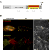
Orthogonal QD targeting to LAP and BirA acceptor peptide (AP) fusion proteins for two-color single molecule imaging. (A) Labeling protocol. Biotinylation of AP was achieved in the secretory pathway by an endoplasmic reticulum-localized BirA (BirA-ER). 10-Br-AMP ligation onto LAP was catalyzed by purified W37ALplA added to the culture media. After rinsing, cells were simultaneously treated with HaloTag-QD605 and streptavidin-QD655. (B) HeLa cultures expressing LAP-LDL receptor and AP-EGF receptor on the same cell (top row), or LAP-neurexin1β and AP-neuroligin1 on neighboring cells (bottom row) were labeled according to the scheme in (A) with 1–10 nM QDs and imaged live. QD605 (yellow) and QD655 (red) were imaged under epifluorescence and total internal reflection fluorescence (TIRF) modes and shown next to DIC images. Scale bars, 10 μm. Movie 1 shows time-lapse TIRF imaging of LAP-neurexin1β and AP-neuroligin1, labeled as in (B).
Figure 4
Figure 4
Measurement of neurexin1β diffusion in the presence vs. absence of neuroligin1 by single-molecule tracking. (A) Left: HeLa cells expressing LAP-neurexin1β were labeled with HaloTag-QD605, then imaged live by TIRF microscopy at 20 Hz. Individual QD tracks from 4 cells were used to calculate diffusion coefficients, plotted here as a histogram. Right: The same experiment, but with AP-neuroligin1 co-expressed. Diffusion coefficients were not significantly different (p = 0.2768). (B) The same experiment as in (A), but with the labels reversed. Neurexin1β was tagged with AP and labeled with streptavidin-QD655. On the right, LAP-neuroligin1 was co-expressed. Again, the diffusion coefficients were not significantly changed by co-expression of neuroligin1 (p = 0.3165). Pink bars indicate interquartile ranges.

References

    1. Pinaud F, Clarke S, Sittner A, Dahan M. Probing Cellular Events, One Quantum Dot at a Time. Nat Methods. 2010;7:275–285. - PubMed
    1. Howarth M, Takao K, Hayashi Y, Ting AY. Targeting Quantum Dots to Surface Proteins in Living Cells with Biotin Ligase. Proc Natl Acad Sci U S A. 2005;102:7583–7588. - PMC - PubMed
    1. Bates IR, Hebert B, Luo Y, Liao J, Bachir AI, Kolin DL, Wiseman PW, Hanrahan JW. Membrane Lateral Diffusion and Capture of CFTR within Transient Confinement Zones. Biophys J. 2006;91:1046–1058. - PMC - PubMed
    1. Sung K, Maloney MT, Yang J, Wu C. A Novel Method for Producing Mono-Biotinylated, Biologically Active Neurotrophic Factors: An Essential Reagent for Single Molecule Study of Axonal Transport. J Neurosci Methods. 2001;200:121–128. - PMC - PubMed
    1. Howarth M, Liu W, Puthenveetil S, Zheng Y, Marshall LF, Schmidt MM, Wittrup KD, Bawendi MG, Ting AY. Monovalent, Reduced-Size Quantum Dots for Imaging Receptors on Living Cells. Nat Methods. 2008;5:397–399. - PMC - PubMed

Publication types