

Immunohistochemical analysis of medullary breast carcinoma autoantigens in different histological types of breast carcinomas
- PMID: 23181716
- PMCID: PMC3533517
- DOI: 10.1186/1746-1596-7-161
Immunohistochemical analysis of medullary breast carcinoma autoantigens in different histological types of breast carcinomas
Abstract
Background: On the past decade a plethora of investigations were directed on identification of molecules involved in breast tumorogenesis, which could represent a powerful tool for monitoring, diagnostics and treatment of this disease. In current study we analyzed six previously identified medullary breast carcinoma autoantigens including LGALS3BP, RAD50, FAM50A, RBPJ, PABPC4, LRRFIP1 with cancer restricted serological profile in different histological types of breast cancer.
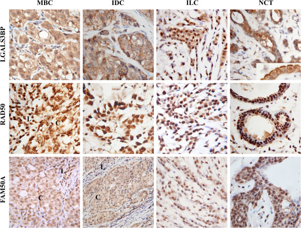
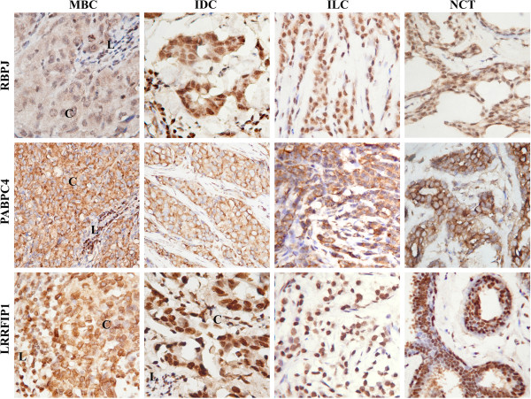
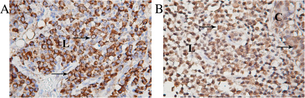
Methods: Semi-quantitative immunohistochemical analysis of 20 tissue samples including medullary breast carcinoma, invasive ductal carcinoma, invasive lobular carcinoma and non-cancerous tissues obtained from patients with fibrocystic disease (each of five) was performed using specifically generated polyclonal antibodies. Differences in expression patterns were evaluated considering percent of positively stained cells, insensitivity of staining and subcellular localization in cells of all tissue samples.
Results: All 6 antigens predominantly expressed in the most cells of all histological types of breast tumors and non-cancerous tissues with slight differences in intensity of staining and subcellular localization. The most significant differences in expression pattern were revealed for RAD50 and LGALS3BP in different histological types of breast cancer and for PABPC4 and FAM50A antigens in immune cells infiltrating breast tumors.
Conclusions: This pilot study made possible to select 4 antigens LGALS3BP, RAD50, PABPC4, and FAM50A as promising candidates for more comprehensive research as potential molecular markers for breast cancer diagnostics and therapy.
Virtual slides: The virtual slides' for this article can be found here: http://www.diagnosticpathology.diagnomx.eu/vs/1860649350796892.
Figures
Similar articles
-
Autoantibody Response to ZRF1 and KRR1 SEREX Antigens in Patients with Breast Tumors of Different Histological Types and Grades.Dis Markers. 2016;2016:5128720. doi: 10.1155/2016/5128720. Epub 2016 Oct 25. Dis Markers. 2016. PMID: 27847402 Free PMC article.
-
Usefulness and limitations of E-cadherin and β-catenin in the classification of breast carcinomas in situ with mixed pattern.Diagn Pathol. 2013 Jul 9;8:114. doi: 10.1186/1746-1596-8-114. Diagn Pathol. 2013. PMID: 23837653 Free PMC article.
-
[Expression of the adhesion molecule CD44v6 in infiltrating ductal carcinomas of the breast is associated with hormone dependence. Our experience with 168 cases].Rev Esp Med Nucl. 2000 Sep;19(5):350-5. doi: 10.1016/s0212-6982(00)71889-1. Rev Esp Med Nucl. 2000. PMID: 11062111 Spanish.
-
Histology and immunophenotype of invasive lobular breast cancer. daily practice and pitfalls.Breast Dis. 2008-2009;30:15-9. doi: 10.3233/BD-2009-0278. Breast Dis. 2008. PMID: 19850991 Review.
-
Breast cancer pathology: the impact of molecular taxonomy on morphological taxonomy.Pathol Int. 2012 May;62(5):295-302. doi: 10.1111/j.1440-1827.2012.02790.x. Epub 2012 Mar 16. Pathol Int. 2012. PMID: 22524656 Review.
Cited by
-
The clinical significance and oncogenic function of LRRFIP1 in pancreatic cancer.Discov Oncol. 2024 Apr 18;15(1):123. doi: 10.1007/s12672-024-00977-3. Discov Oncol. 2024. PMID: 38634978 Free PMC article.
-
Upregulation of FAM50A promotes cancer development.Med Oncol. 2023 Jul 1;40(8):217. doi: 10.1007/s12032-023-02072-z. Med Oncol. 2023. PMID: 37393403
-
Proto-Oncogene FAM50A Can Regulate the Immune Microenvironment and Development of Hepatocellular Carcinoma In Vitro and In Vivo.Int J Mol Sci. 2023 Feb 6;24(4):3217. doi: 10.3390/ijms24043217. Int J Mol Sci. 2023. PMID: 36834630 Free PMC article.
-
Aortic α-smooth muscle actin expressions in aortic disorders and coronary artery disease: An immunohistochemical study.Anatol J Cardiol. 2018 Jan;19(1):11-16. doi: 10.14744/AnatolJCardiol.2017.7839. Anatol J Cardiol. 2018. PMID: 29339694 Free PMC article.
-
Galectin-3 Binding Protein Secreted by Breast Cancer Cells Inhibits Monocyte-Derived Fibrocyte Differentiation.J Immunol. 2015 Aug 15;195(4):1858-67. doi: 10.4049/jimmunol.1500365. Epub 2015 Jul 1. J Immunol. 2015. PMID: 26136428 Free PMC article.
References
-
- Chen YT. Identification of human tumor antigens by serological expression cloning: an online review on SEREX. Cancer Immunol. 2004. http://www.cance-rimmunity.org/SEREX/
Publication types
MeSH terms
Substances
LinkOut - more resources
Full Text Sources
Medical
Research Materials
Miscellaneous
