. 2013 Feb 15;450(1):141-8.
doi: 10.1042/BJ20121434.

Role of Helicobacter pylori methionine sulfoxide reductase in urease maturation

Affiliations

Role of Helicobacter pylori methionine sulfoxide reductase in urease maturation

Lisa G Kuhns et al. Biochem J. .

Abstract

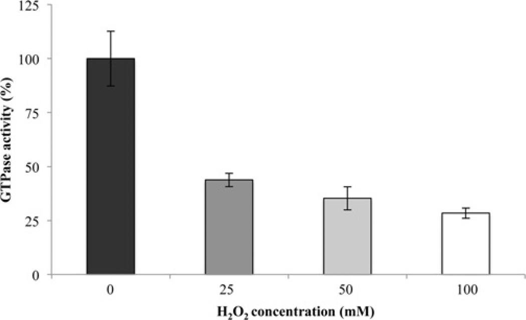
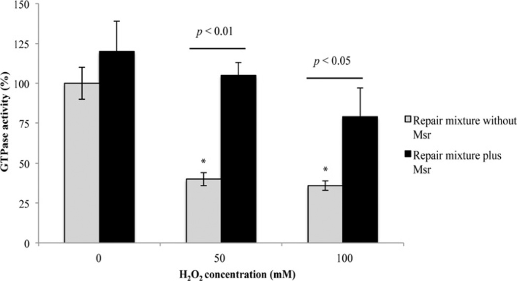
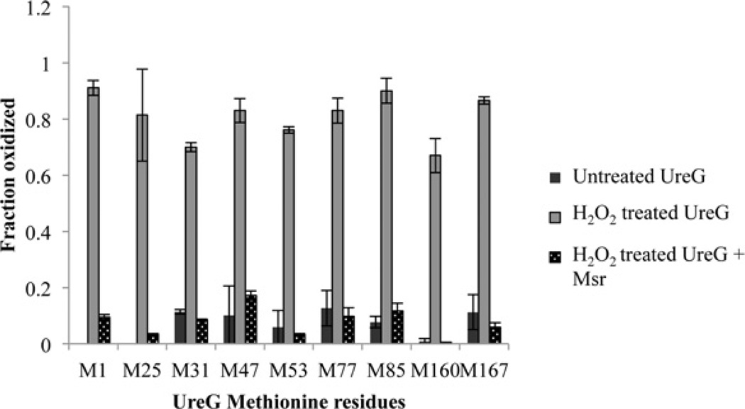
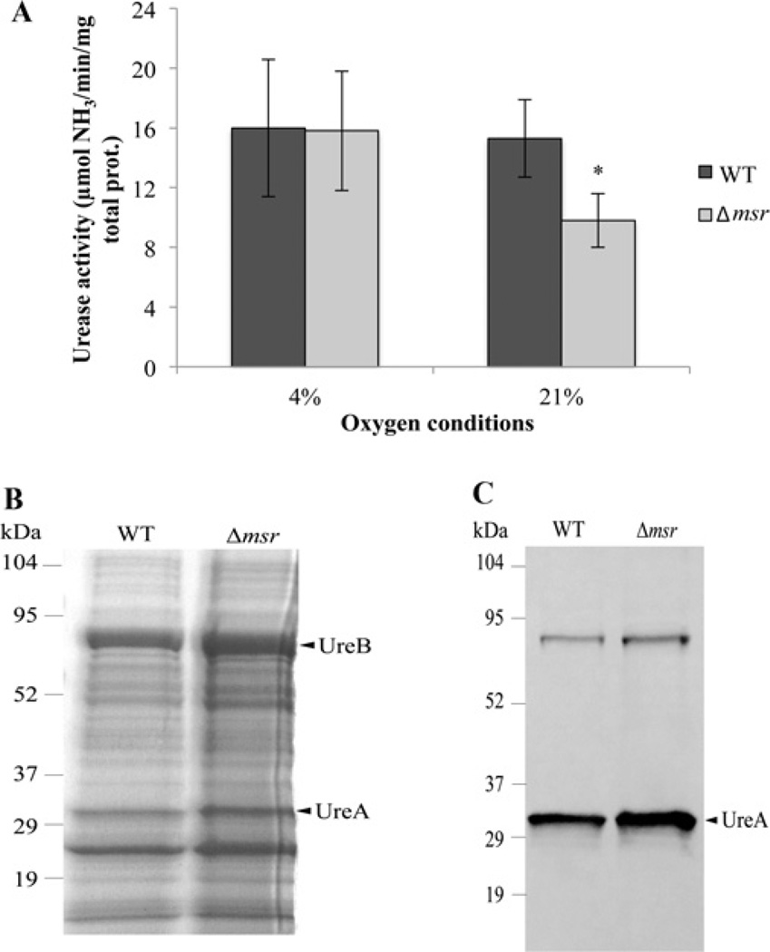
The persistence of the gastric pathogen Helicobacter pylori is due in part to urease and Msr (methionine sulfoxide reductase). Upon exposure to relatively mild (21% partial pressure of O2) oxidative stress, a Δmsr mutant showed both decreased urease specific activity in cell-free extracts and decreased nickel associated with the partially purified urease fraction as compared with the parent strain, yet urease apoprotein levels were the same for the Δmsr and wild-type extracts. Urease activity of the Δmsr mutant was not significantly different from the wild-type upon non-stress microaerobic incubation of strains. Urease maturation occurs through nickel mobilization via a suite of known accessory proteins, one being the GTPase UreG. Treatment of UreG with H2O2 resulted in oxidation of MS-identified methionine residues and loss of up to 70% of its GTPase activity. Incubation of pure H2O2-treated UreG with Msr led to reductive repair of nine methionine residues and recovery of up to full enzyme activity. Binding of Msr to both oxidized and non-oxidized UreG was observed by cross-linking. Therefore we conclude Msr aids the survival of H. pylori in part by ensuring continual UreG-mediated urease maturation under stress conditions.


Figures

Figure 1
Figure 1. SDS/PAGE of purified UreG
Lane 1, molecular mass marker with values indicated to the left in kDa; lane 2, cell extract from non-induced E. coli BL21(DE3)-RIL harbouring pET21b-ureG; lane 3, cell extract from IPTG-induced E. coli BL21(DE3)-RIL harbouring pET21b-ureG; lane 4, purified UreG after Ni-NTA extraction; lane 5, concentrated pure UreG. The arrowhead to the right indicates UreG.
Figure 2
Figure 2. Interaction between UreG and Msr identified by biotin transfer
Sulfo-SBED-conjugated UreG or 50 mMH2O2-oxidized UreG was incubated with Msr, lysozyme or alone and then the mixture was subjected to UV-cross-linking. Samples were taken before and after exposure to UV light. The conjugated samples were reduced with 0.5 M DTT for label transfer and the proteins were resolved via SDS/PAGE, transferred on to a nitrocellulose membrane, and probed with streptavidin–HRP. Lane 1, UreG; lane 2, oxidized UreG; lane 3, UreG and lysozyme; lane 4, oxidized UreG and lysozyme; lane 5, UreG and Msr; lane 6, oxidized UreG and Msr; lane 7, UreG; lane 8, oxidized UreG; lane 9, UreG and Msr; lane 10, oxidized UreG and Msr. Samples in lanes 7–10 were not exposed to UV light. The molecular masses are displayed to the left with values in kDa. The arrowheads to the right indicate Msr or UreG. UreG is ~22 kDa and Msr is ~42 kDa.
Figure 3
Figure 3. H2O2 inactivation of UreG GTPase activity
Purified UreG (6 µM) was incubated with various concentrations of H2O2 (0, 25, 50 or 100 mM) for 3 h. Excess oxidant was removed via overnight dialysis. GTPase activities were measured using a colorimetric assay to detect the release of Pi and are presented as percentage activity of the untreated sample. The untreated sample (100%) is 0.186 µmol Pi/min per mg of UreG. Each concentration is statistically significantly less than every higher concentration shown in the Figure at P<0.05. Results are means ±S.D. (n = 8, two independent experiments were each sampled four times).
Figure 4
Figure 4. Msr repair of H2O2-damaged UreG
H2O2-treated UreG (6 µM) was incubated with equimolar amounts of Msr or buffer, along with the Msr repair components (400 µM NADPH, 5 µM Trx and 100 nM TrxR) at 37°C for 1 h. The samples were then assayed for GTPase activity spectrophotometrically. UreG GTPase activity of the untreated sample without the addition of Msr is considered as 100% and is 0.136 µmol of Pi/min per mg of UreG. Results are means ±S.D. (n = 6, three independent experiments sampled in duplicate).
Figure 5
Figure 5. MS/MS identification of methionine residues after oxidation and repair of UreG
UreG was incubated with buffer or 50 mM H2O2 in buffer for 3 h. Excess H2O2 was removed via overnight dialysis. Following dialysis, oxidized UreG samples were incubated with or without Msr in the presence of DTT at 37°C for 1 h. DTT alone did not result in the repair of any methionine residues. No oxidized Met1 or Met25 could be detected in the untreated sample. Samples were digested with Asp-N and methionine residues were identified and quantified by LC–MS/MS.
Figure 6
Figure 6. Urease activity and expression after oxidant stress
(A) Urease activity after exposure to oxidative stress. H. pylori SS1 wild-type (WT) and Δmsr strains were exposed to 21% O2 (air) for 2 h or left at 4% O2 as a control. Cells were then lysed by sonication and urease activity was measured in cell-free extracts. Results are means ±S.D (n = 12, based on four independent experiments each sampled in triplicate). *P < 0.01. (B) SDS/PAGE analysis of cell-free extract from SS1 wild-type (9 µg) and Δmsr (10 µg) after exposure to 21%O2. (C) Immunoblot analysis of SS1 wild-type and Δmsr cell-free extract after exposure to 21% O2. Whole-cell extracts were resolved via SDS/PAGE, transferred on to a nitrocellulose membrane and blotted with anti-UreA antibodies. Molecular masses in kDa are indicated to the left-hand side of (B) and (C).

Similar articles

Cited by

References

    1. Marshall BJ, Warren JR. Unidentified curved bacilli in the stomach of patients with gastritis and peptic ulceration. Lancet. 1984;1:1311–1315. - PubMed
    1. Blaser MJ, Atherton JC. Helicobacter pylori persistence: biology and disease. J. Clin. Invest. 2004;113:321–333. - PMC - PubMed
    1. Dunn BE, Cohen H, Blaser MJ. Helicobacter pylori. Clin. Microbiol. Rev. 1997;10:720–741. - PMC - PubMed
    1. Bagchi D, Bhattacharya G, Stohs SJ. Production of reactive oxygen species by gastric cells in association with Helicobacter pylori. Free Radical Res. 1996;24:439–450. - PubMed
    1. Ramarao N, Gray-Owen SD, Meyer TF. Helicobacter pylori induces but survives the extracellular release of oxygen radicals from professional phagocytes using its catalase activity. Mol. Microbiol. 2000;38:103–113. - PubMed

Publication types

MeSH terms

LinkOut - more resources