. 2013 Jan 18;33(1):165-73.
doi: 10.1042/BSR20120112.

Characterization of the bipartite degron that regulates ubiquitin-independent degradation of thymidylate synthase

Affiliations

Characterization of the bipartite degron that regulates ubiquitin-independent degradation of thymidylate synthase

Karen W Barbour et al. Biosci Rep. .

Abstract

TS (thymidylate synthase) is a key enzyme in the de novo biosynthesis of dTMP, and is indispensable for DNA replication. Previous studies have shown that intracellular degradation of the human enzyme [hTS (human thymidylate synthase)] is mediated by the 26S proteasome, and occurs in a ubiquitin-independent manner. Degradation of hTS is governed by a degron that is located at the polypeptide's N-terminus that is capable of promoting the destabilization of heterologous proteins to which it is attached. The hTS degron is bipartite, consisting of two subdomains: an IDR (intrinsically disordered region) that is highly divergent among mammalian species, followed by a conserved amphipathic α-helix (designated hA). In the present report, we have characterized the structure and function of the hTS degron in more detail. We have conducted a bioinformatic analysis of interspecies sequence variation exhibited by the IDR, and find that its hypervariability is not due to diversifying (or positive) selection; rather, it has been subjected to purifying (or negative) selection, although the intensity of such selection is relaxed or weakened compared with that exerted on the rest of the molecule. In addition, we have verified that both subdomains of the hTS degron are required for full activity. Furthermore, their co-operation does not necessitate that they are juxtaposed, but is maintained when they are physically separated. Finally, we have identified a 'cryptic' degron at the C-terminus of hTS, which is activated by the N-terminal degron and appears to function only under certain circumstances; its role in TS metabolism is not known.


Figures
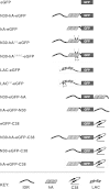
Figure 1
Figure 1. Constructs used in the present study
The eGFP fusion constructs utilized in the current study are shown. Schematic depictions of the various segments are indicated in the KEY at the bottom, including the disordered region (IDR), the amphipathic α-helix (hA) and the C-terminal element (C38) of hTS, as well as the N-terminal region of the E. coli lac repressor (LAC).
Figure 2
Figure 2. Cumulative behaviour of synonymous and non-synonymous substitutions along the coding region of TS mRNA
Accumulation of amino acid substitutions within the TS polypeptide for the 16 mammalian species, as determined by SNAP, is plotted along the coding region of the mRNA (see the Experimental section). Codon numbering refers to hTS. The entire coding region (313 codons) is shown in (A), whereas a closer view of the first 80 codons is shown in (B). The plot for synonymous substitutions is in red, while that for non-synonymous substitutions is in green. The black line shows accumulation of insertions/deletions. The IDR is indicated by a double-arrowed dashed line (<—>).
Figure 3
Figure 3. Rates of accumulation of synonymous and non-synonymous substitutions along the TS mRNA
Slopes of the plots in Figure 2 were determined by linear regression. Codons 1–27 represent the IDR of hTS, while codons 28-313 represent the rest of the polypeptide. Bars indicate values±S.E.M. P values are shown for various comparisons, as indicated by the brackets.
Figure 4
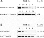
Figure 4. hA is inactive on its own as a degron
The indicated plasmid constructs (see the text and Figure 1) were stably transfected into cell line RJK88.13 and treated with CHX. Decreases in protein concentrations were monitored over time by Western blotting (see the Experimental section for details). A representative blot is shown for each construct. The half-lives are shown to the right of the corresponding blots, and are presented as means±S.D.
Figure 5
Figure 5. Predicted impact of amino acid substitutions on the helical propensity of hA segment
The helical propensity of the region spanning residues 24–50 of wild-type hTS and mutants I39/I42, P40/P41 and I39/P40/P41/I42 were determined using PSIPRED [27]. For each molecule, the amino acid sequence is shown; targeted substitutions within mutated segments are indicated in red. The predicted secondary structure is shown below each sequence; black lines indicate coiled regions, magenta cylinders indicate α-helices and yellow arrows indicate β-strands. The blue bars at the bottom show the relative confidence for the predictions at each position along the sequence, with the height and depth of blue indicating greater confidence. The predictor accurately identified hA of wild-type hTS, known from X-ray crystallographic studies [4,5].
Figure 6
Figure 6. Degron activity requires a helical segment
The indicated plasmid constructs were stably transfected into cell line RJK88.13 and treated with CHX (see the text and Figure 1). Decreases in protein concentrations over time were monitored by Western blotting (see the Experimental section for details). A representative blot is shown for each construct. The half-lives are shown to the right of the corresponding blots, and are presented as means±S.D.
Figure 7
Figure 7. The IDR and hA co-operate when physically separated
Plasmid constructs were stably transfected into cell line RJK88.13 and treated with CHX (see the text and Figure 1). Decreases in protein concentrations over time were monitored by Western blotting (see the Experimental section for details). A representative blot is shown for each construct. The half-lives are shown to the right of the corresponding blots, and are presented as means±S.D.
Figure 8
Figure 8. A ‘cryptic’ degron at the C-terminus of hTS
Plasmids expressing (see the text and Figure 1) were stably transfected into cell line RJK88.13 and treated with CHX. Decreases in protein concentrations over time were monitored by Western blotting (see the Experimental section for details). A representative blot is shown for each construct. The half-lives are shown to the right of the corresponding blots, and are presented as means±S.D.

Similar articles

Cited by

References

    1. Berger F. G., Berger S. H. Thymidylate synthase as a chemotherapeutic drug target: where are we after fifty years? Cancer Biol. Ther. 2006;5:1238–1241. - PubMed
    1. Carreras C. W., Santi D. V. The catalytic mechanism and structure of thymidylate synthase. Annu. Rev. Biochem. 1995;64:721–762. - PubMed
    1. Hardy L. W., Finer-Moore J. S., Montfort W. R., Jones M. O., Santi D. V., Stroud R. M. Atomic structure of thymidylate synthase: target for rational drug design. Science. 1987;235:448–455. - PubMed
    1. Phan J., Steadman D. J., Koli S., Ding W. C., Minor W., Dunlap R. B., Berger S. H., Lebioda L. Structure of human thymidylate synthase suggests advantages of chemotherapy with noncompetitive inhibitors. J. Biol. Chem. 2001;276:14170–14177. - PubMed
    1. Schiffer C. A., Clifton I. J., Davisson V. J., Santi D. V., Stroud R. M. Crystal structure of human thymidylate synthase: a structural mechanism for guiding substrates into the active site. Biochemistry. 1995;34:16279–16287. - PubMed

Publication types

MeSH terms