

Fatigue in muscular dystrophies
- PMID: 23182642
- PMCID: PMC3526799
- DOI: 10.1016/j.nmd.2012.10.010
Fatigue in muscular dystrophies
Abstract
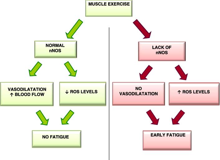
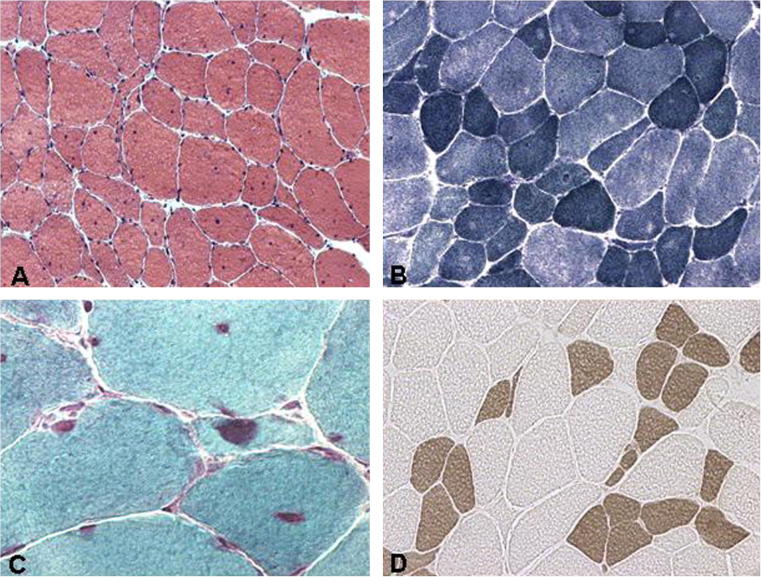
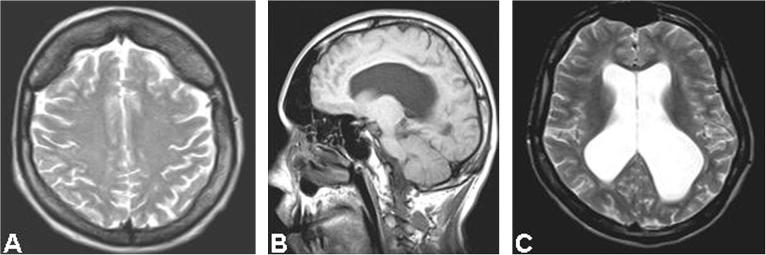
Fatigue is a frequent complaint in muscular dystrophies but it is yet not well defined or studied. We have examined the issue of muscle fatigue in a series of molecularly defined muscular dystrophies. A greater fatigability is seen in muscular dystrophy patients and can be an acute or chronic status. In Duchenne Muscular Dystrophy and beta-sarcoglycanopathy besides the alteration of dystrophin and/or sarcoglycan complex, a neuronal nitric oxide synthase depletion is frequently found and might correlate with post-exercise fatigability as well as with cardiac involvement. Therefore, it might be an important modulating factor of the severity of myopathy. In myotonic dystrophy, fatigue is a common complaint: muscle is involved and type 1 atrophy is a frequent feature; brain involvement and depressed mood might likely explain the extent of fatigue and daytime sleepiness commonly observed in these patients. Furthermore, in our observation in a series of 24 cases, muscle and brain can be independently involved in DM1 patients. These observations have profound impact on the type of physical therapy to be prescribed in such patients.
Copyright © 2012 Elsevier B.V. All rights reserved.
Figures
References
-
- Feasson L., Camdessanche J.P., El Mandhi L., Calmels P., Millet G.Y. Fatigue et affection neuromusculaire. Ann Readapt Med Phys. 2006;49:289–300. - PubMed
-
- Stramare R., Beltrame V., Dal Borgo R. MRI in the assessment of muscular pathology: a comparison between limb-girdle muscular dystrophies, hyaline body myopathies and myotonic dystrophies. Radiol Med. 2010;115:585–599. - PubMed
Publication types
MeSH terms
Substances
Grants and funding
LinkOut - more resources
Full Text Sources
Other Literature Sources
Medical
