

In vitro growth factor-induced bio engineering of mature articular cartilage
- PMID: 23182922
- PMCID: PMC3543901
- DOI: 10.1016/j.biomaterials.2012.09.076
In vitro growth factor-induced bio engineering of mature articular cartilage
Abstract
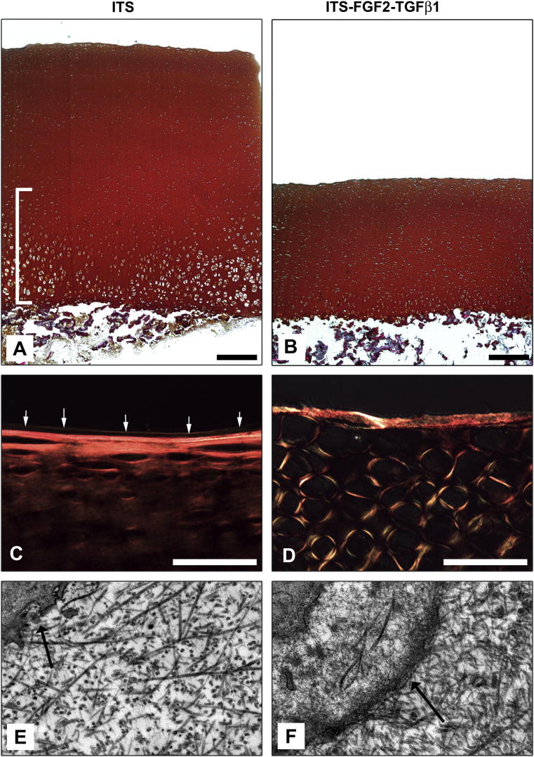
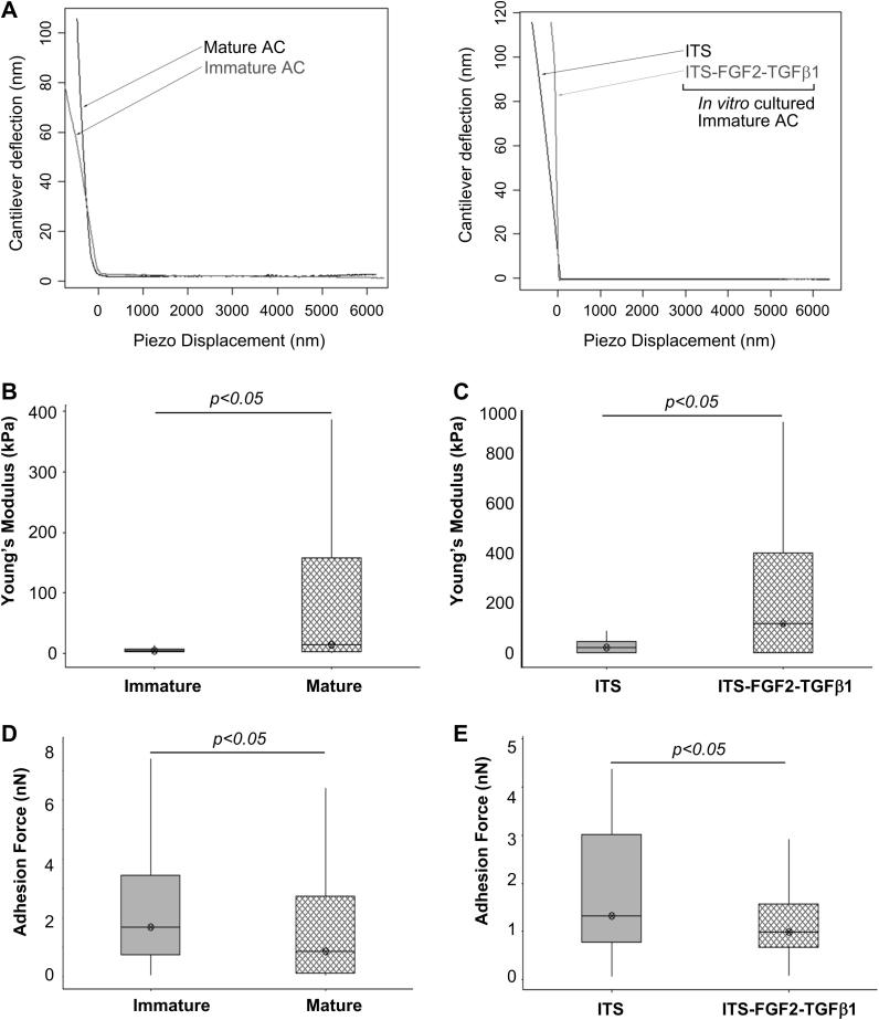
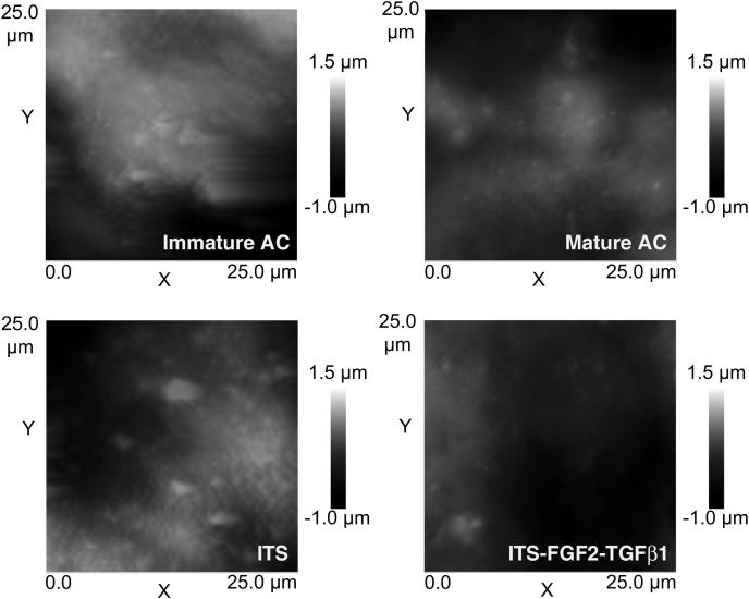
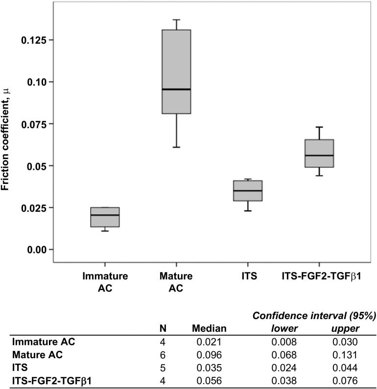
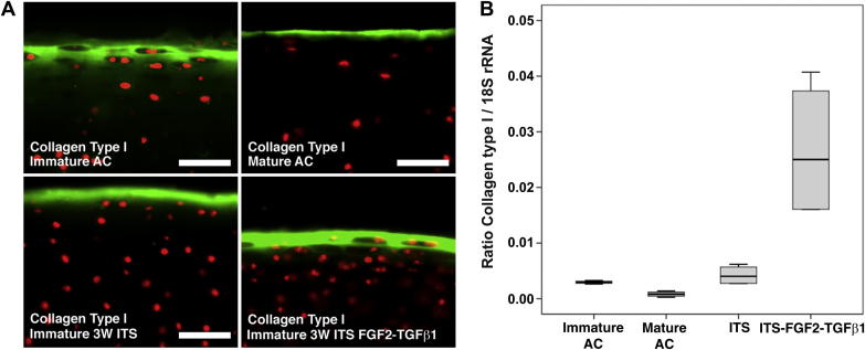
Articular cartilage maturation is the postnatal development process that adapts joint surfaces to their site-specific biomechanical demands. Maturation involves gross morphological changes that occur through a process of synchronised growth and resorption of cartilage and generally ends at sexual maturity. The inability to induce maturation in biomaterial constructs designed for cartilage repair has been cited as a major cause for their failure in producing persistent cell-based repair of joint lesions. The combination of growth factors FGF2 and TGFβ1 induces accelerated articular cartilage maturation in vitro such that many molecular and morphological characteristics of tissue maturation are observable. We hypothesised that experimental growth factor-induced maturation of immature cartilage would result in a biophysical and biochemical composition consistent with a mature phenotype. Using native immature and mature cartilage as reference, we observed that growth factor-treated immature cartilages displayed increased nano-compressive stiffness, decreased surface adhesion, decreased water content, increased collagen content and smoother surfaces, correlating with a convergence to the mature cartilage phenotype. Furthermore, increased gene expression of surface structural protein collagen type I in growth factor-treated explants compared to reference cartilages demonstrates that they are still in the dynamic phase of the postnatal developmental transition. These data provide a basis for understanding the regulation of postnatal maturation of articular cartilage and the application of growth factor-induced maturation in vitro and in vivo in order to repair and regenerate cartilage defects.
Crown Copyright © 2012. Published by Elsevier Ltd. All rights reserved.
Figures
Similar articles
-
Fibroblast growth factor 2 and transforming growth factor β1 induce precocious maturation of articular cartilage.Arthritis Rheum. 2011 Nov;63(11):3417-27. doi: 10.1002/art.30543. Arthritis Rheum. 2011. PMID: 21769844
-
Differential regulation of immature articular cartilage compressive moduli and Poisson's ratios by in vitro stimulation with IGF-1 and TGF-beta1.J Biomech. 2010 Sep 17;43(13):2501-7. doi: 10.1016/j.jbiomech.2010.05.022. Epub 2010 Jun 8. J Biomech. 2010. PMID: 20570267 Free PMC article.
-
Expansion of human articular chondrocytes and formation of tissue-engineered cartilage: a step towards exploring a potential use of matrix-induced cell therapy.Tissue Cell. 2010 Oct;42(5):282-92. doi: 10.1016/j.tice.2010.07.002. Tissue Cell. 2010. PMID: 20810142
-
Bioengineering cartilage growth, maturation, and form.Pediatr Res. 2008 May;63(5):527-34. doi: 10.1203/PDR.0b013e31816b4fe5. Pediatr Res. 2008. PMID: 18427298 Review.
-
Animal models for cartilage repair.J Biol Regul Homeost Agents. 2018 Nov-Dec;32(6 Suppl. 1):105-116. J Biol Regul Homeost Agents. 2018. PMID: 30644290 Review.
Cited by
-
Over-Production of Therapeutic Growth Factors for Articular Cartilage Regeneration by Protein Production Platforms and Protein Packaging Cell Lines.Biology (Basel). 2020 Oct 9;9(10):330. doi: 10.3390/biology9100330. Biology (Basel). 2020. PMID: 33050357 Free PMC article. Review.
-
An injectable, in situ forming type II collagen/hyaluronic acid hydrogel vehicle for chondrocyte delivery in cartilage tissue engineering.Drug Deliv Transl Res. 2014 Apr;4(2):149-58. doi: 10.1007/s13346-013-0188-1. Drug Deliv Transl Res. 2014. PMID: 25786729
-
Advances in regenerative orthopedics.Mayo Clin Proc. 2013 Nov;88(11):1323-39. doi: 10.1016/j.mayocp.2013.04.027. Mayo Clin Proc. 2013. PMID: 24182709 Free PMC article.
-
The regenerative role of adipose-derived stem cells (ADSC) in plastic and reconstructive surgery.Int Wound J. 2017 Feb;14(1):112-124. doi: 10.1111/iwj.12569. Epub 2016 Feb 1. Int Wound J. 2017. PMID: 26833722 Free PMC article. Review.
-
The bio in the ink: cartilage regeneration with bioprintable hydrogels and articular cartilage-derived progenitor cells.Acta Biomater. 2017 Oct 1;61:41-53. doi: 10.1016/j.actbio.2017.08.005. Epub 2017 Aug 4. Acta Biomater. 2017. PMID: 28782725 Free PMC article.
References
-
- Mow V.C., Ratcliffe A., Poole A.R. Cartilage and diarthrodial joints as paradigms for hierarchical materials and structures. Biomaterials. 1992;13(2):67–97. - PubMed
-
- Khan I.M., Gilbert S.J., Singhrao S.K., Duance V.C., Archer C.W. Cartilage integration: evaluation of the reasons for failure of integration during cartilage repair. A review. Eur Cell Mater. 2008;16:26–39. - PubMed
-
- Namba R.S., Meuli M., Sullivan K.M., Le A.X., Adzick N.S. Spontaneous repair of superficial defects in articular cartilage in a fetal lamb model. J Bone Joint Surg Am. 1998;80(1):4–10. - PubMed
-
- Shapiro F., Koide S., Glimcher M.J. Cell origin and differentiation in the repair of full-thickness defects of articular cartilage. J Bone Joint Surg Am. 1993;75(4):532–553. - PubMed
-
- Grande D.A., Pitman M.I., Peterson L., Menche D., Klein M. The repair of experimentally produced defects in rabbit articular cartilage by autologous chondrocyte transplantation. J Orthop Res. 1989;7(2):208–218. - PubMed
Publication types
MeSH terms
Substances
Grants and funding
LinkOut - more resources
Full Text Sources
Other Literature Sources
